3ZQC | pdb_00003zqc

Structure of the Trichomonas vaginalis Myb3 DNA-binding domain bound to a promoter sequence reveals a unique C-terminal beta-hairpin conformation


Experimental Data Snapshot

  • Method: X-RAY DIFFRACTION
  • Resolution: 2.90 Å
  • R-Value Free: 
    0.273 (Depositor), 0.286 (DCC) 
  • R-Value Work: 
    0.218 (Depositor), 0.227 (DCC) 
  • R-Value Observed: 
    0.218 (Depositor) 

Starting Model: experimental
View more details

wwPDB Validation 3D Report Full Report

Validation slider image for 3ZQC

This is version 1.1 of the entry. See complete history

Literature

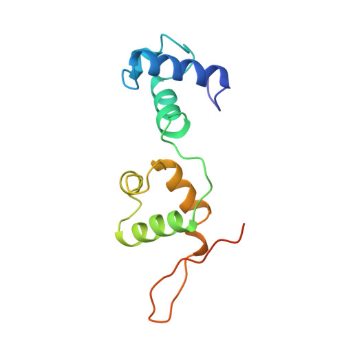
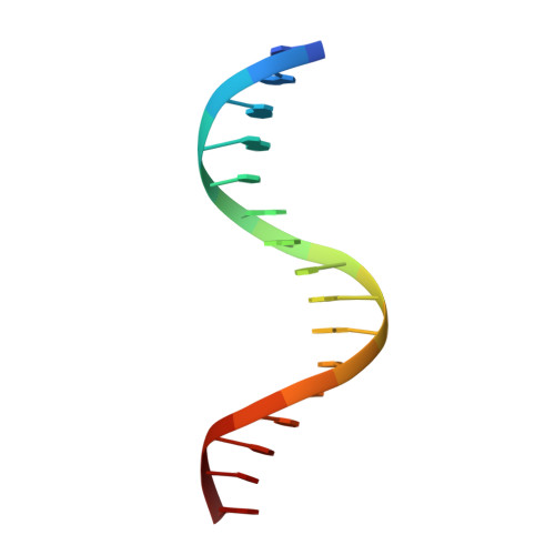
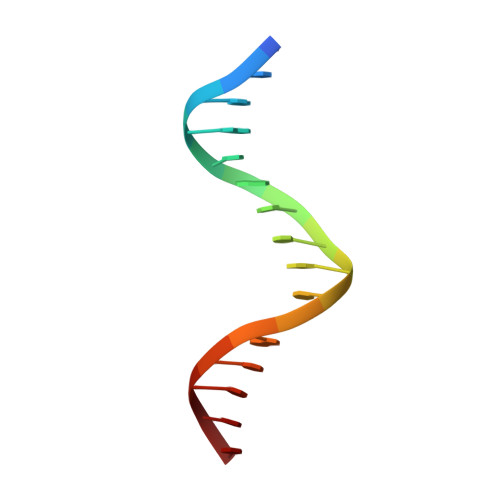
Structure of the Trichomonas Vaginalis Myb3 DNA-Binding Domain Bound to a Promoter Sequence Reveals a Unique C-Terminal Beta-Hairpin Conformation.

Wei, S.-Y.Lou, Y.-C.Tsai, J.-Y.Ho, M.R.Chou, C.C.Rajasekaran, M.Hsu, H.-M.Tai, J.-H.Hsiao, C.-D.Chen, C.

(2012) Nucleic Acids Res 40: 449

  • DOI: https://doi.org/10.1093/nar/gkr707
  • Primary Citation Related Structures: 
    3ZQC

  • PubMed Abstract: 

    Trichomonas vaginalis Myb3 transcription factor (tvMyb3) recognizes the MRE-1 promoter sequence and regulates ap65-1 gene, which encodes a hydrogenosomal malic enzyme that may play a role in the cytoadherence of the parasite. Here, we identified tvMyb3(53-180) as the essential fragment for DNA recognition and report the crystal structure of tvMyb3(53-180) bound to MRE-1 DNA. The N-terminal fragment adopts the classical conformation of an Myb DNA-binding domain, with the third helices of R2 and R3 motifs intercalating in the major groove of DNA. The C-terminal extension forms a β-hairpin followed by a flexible tail, which is stabilized by several interactions with the R3 motif and is not observed in other Myb proteins. Interestingly, this unique C-terminal fragment does not stably connect with DNA in the complex structure but is involved in DNA binding, as demonstrated by NMR chemical shift perturbation, (1)H-(15)N heteronuclear-nuclear Overhauser effect and intermolecular paramagnetic relaxation enhancement. Site-directed mutagenesis also revealed that this C-terminal fragment is crucial for DNA binding, especially the residue Arg(153) and the fragment K(170)KRK(173). We provide a structural basis for MRE-1 DNA recognition and suggest a possible post-translational regulation of tvMyb3 protein.


  • Organizational Affiliation
    • Institute of Biomedical Sciences, Academia Sinica, Taipei 115, Taiwan, ROC.

Macromolecule Content 

  • Total Structure Weight: 100.69 kDa 
  • Atom Count: 6,627 
  • Modeled Residue Count: 605 
  • Deposited Residue Count: 652 
  • Unique protein chains: 1
  • Unique nucleic acid chains: 2

Macromolecules


Find similar proteins by:|  3D Structure
Entity ID: 1
MoleculeChains  Sequence LengthOrganismDetailsImage
MYB3
A, D, G, J
131Trichomonas vaginalisMutation(s): 0 
UniProt
Find proteins for A2D9X4 (Trichomonas vaginalis (strain ATCC PRA-98 / G3))
Explore A2D9X4 
Go to UniProtKB:  A2D9X4
Entity Groups
Sequence Clusters30% Identity50% Identity70% Identity90% Identity95% Identity100% Identity
UniProt GroupA2D9X4
Sequence Annotations
Expand
Reference Sequence
Find similar nucleic acids by:  Sequence
Entity ID: 2
MoleculeChains LengthOrganismImage
MRE-1
B, E, H, K
16Trichomonas vaginalis
Sequence Annotations
Expand
Reference Sequence
Find similar nucleic acids by:  Sequence
Entity ID: 3
MoleculeChains LengthOrganismImage
MRE-1
C, F, I, L
16Trichomonas vaginalis
Sequence Annotations
Expand
Reference Sequence

Experimental Data & Validation

Experimental Data

  • Method: X-RAY DIFFRACTION
  • Resolution: 2.90 Å
  • R-Value Free:  0.273 (Depositor), 0.286 (DCC) 
  • R-Value Work:  0.218 (Depositor), 0.227 (DCC) 
  • R-Value Observed: 0.218 (Depositor) 
Space Group: P 1
Unit Cell:
Length ( Å )Angle ( ˚ )
a = 45.81α = 94.68
b = 71.77β = 97.84
c = 87.82γ = 99.28
Software Package:
Software NamePurpose
CNSrefinement
HKL-2000data reduction
HKL-2000data scaling
MOLREPphasing

Structure Validation

View Full Validation Report


Entry History 

Deposition Data

Revision History  (Full details and data files)

  • Version 1.0: 2012-04-18
    Type: Initial release
  • Version 1.1: 2023-12-20
    Changes: Data collection, Database references, Other, Refinement description