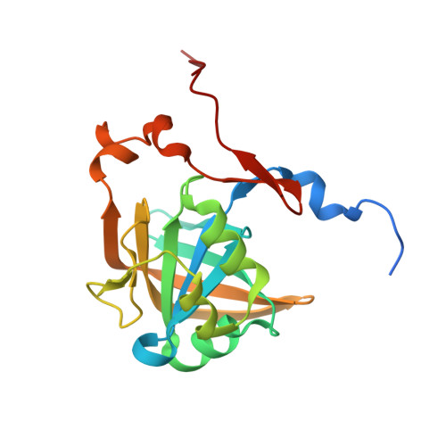
3ZOC

Crystal structure of FMN-binding protein (NP_142786.1) from Pyrococcus horikoshii with bound p-hydroxybenzaldehyde


Experimental Data Snapshot

  • Method: X-RAY DIFFRACTION
  • Resolution: 2.10 Å
  • R-Value Free: 0.269 
  • R-Value Work: 0.242 
  • R-Value Observed: 0.243 

Starting Model: experimental
View more details

wwPDB Validation   3D Report Full Report


Ligand Structure Quality Assessment 


This is version 1.3 of the entry. See complete history


Literature

Identification of Promiscuous Ene-Reductase Activity by Mining Structural Databases Using Active Site Constellations.

Steinkellner, G.Gruber, C.C.Pavkov-Keller, T.Binter, A.Steiner, K.Winkler, C.Lyskowski, A.Schwamberger, O.Oberer, M.Schwab, H.Faber, K.Macheroux, P.Gruber, K.

(2014) Nat Commun 5: 4150

  • DOI: https://doi.org/10.1038/ncomms5150
  • Primary Citation of Related Structures:  
    3ZOC, 3ZOD, 3ZOE, 3ZOF, 3ZOG, 3ZOH

  • PubMed Abstract: 

    The exploitation of catalytic promiscuity and the application of de novo design have recently opened the access to novel, non-natural enzymatic activities. Here we describe a structural bioinformatic method for predicting catalytic activities of enzymes based on three-dimensional constellations of functional groups in active sites ('catalophores'). As a proof-of-concept we identify two enzymes with predicted promiscuous ene-reductase activity (reduction of activated C-C double bonds) and compare them with known ene-reductases, that is, members of the Old Yellow Enzyme family. Despite completely different amino acid sequences, overall structures and protein folds, high-resolution crystal structures reveal equivalent binding modes of typical Old Yellow Enzyme substrates and ligands. Biochemical and biocatalytic data show that the two enzymes indeed possess ene-reductase activity and reveal an inverted stereopreference compared with Old Yellow Enzymes for some substrates. This method could thus be a tool for the identification of viable starting points for the development and engineering of novel biocatalysts.


  • Organizational Affiliation

    1] ACIB GmbH, Petersgasse 14, 8010 Graz, Austria [2].


Macromolecules
Find similar proteins by:  (by identity cutoff)  |  3D Structure
Entity ID: 1
MoleculeChains Sequence LengthOrganismDetailsImage
FMN-BINDING PROTEIN191Pyrococcus horikoshiiMutation(s): 0 
UniProt
Find proteins for O58586 (Pyrococcus horikoshii (strain ATCC 700860 / DSM 12428 / JCM 9974 / NBRC 100139 / OT-3))
Explore O58586 
Go to UniProtKB:  O58586
Entity Groups  
Sequence Clusters30% Identity50% Identity70% Identity90% Identity95% Identity100% Identity
UniProt GroupO58586
Sequence Annotations
Expand
  • Reference Sequence
Experimental Data & Validation

Experimental Data

  • Method: X-RAY DIFFRACTION
  • Resolution: 2.10 Å
  • R-Value Free: 0.269 
  • R-Value Work: 0.242 
  • R-Value Observed: 0.243 
  • Space Group: P 61 2 2
Unit Cell:
Length ( Å )Angle ( ˚ )
a = 46.321α = 90
b = 46.321β = 90
c = 268.77γ = 120
Software Package:
Software NamePurpose
REFMACrefinement
XDSdata reduction
PHASERphasing

Structure Validation

View Full Validation Report


Ligand Structure Quality Assessment 


Entry History 

Deposition Data

Revision History  (Full details and data files)

  • Version 1.0: 2014-05-14
    Type: Initial release
  • Version 1.1: 2014-07-02
    Changes: Database references
  • Version 1.2: 2019-03-06
    Changes: Data collection, Experimental preparation, Other
  • Version 1.3: 2023-12-20
    Changes: Data collection, Database references, Derived calculations, Other, Refinement description