3ZKR | pdb_00003zkr

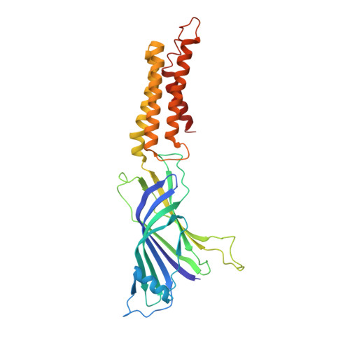
X-ray structure of a pentameric ligand gated ion channel from Erwinia chrysanthemi (ELIC) in complex with bromoform


Experimental Data Snapshot

  • Method: X-RAY DIFFRACTION
  • Resolution: 3.65 Å
  • R-Value Free: 
    0.264 (Depositor), 0.267 (DCC) 
  • R-Value Work: 
    0.229 (Depositor), 0.234 (DCC) 
  • R-Value Observed: 
    0.231 (Depositor) 

wwPDB Validation 3D Report Full Report

Validation slider image for 3ZKR

This is version 1.3 of the entry. See complete history. 

Literature

Multisite Binding of a General Anesthetic to the Prokaryotic Pentameric Erwinia Chrysanthemi Ligand-Gated Ion Channel (Elic).

Spurny, R., Billen, B., Howard, R.J., Brams, M., Debaveye, S., Price, K.L., Weston, D.A., Strelkov, S.V., Tytgat, J., Bertrand, S., Bertrand, D., Lummis, S.C.R., Ulens, C.

(2013) J Biol Chem 288: 8355

  • DOI: https://doi.org/10.1074/jbc.M112.424507
  • Primary Citation Related Structures: 
    3ZKR

  • PubMed Abstract: 

    Pentameric ligand-gated ion channels (pLGICs), such as nicotinic acetylcholine, glycine, γ-aminobutyric acid GABA(A/C) receptors, and the Gloeobacter violaceus ligand-gated ion channel (GLIC), are receptors that contain multiple allosteric binding sites for a variety of therapeutics, including general anesthetics. Here, we report the x-ray crystal structure of the Erwinia chrysanthemi ligand-gated ion channel (ELIC) in complex with a derivative of chloroform, which reveals important features of anesthetic recognition, involving multiple binding at three different sites. One site is located in the channel pore and equates with a noncompetitive inhibitor site found in many pLGICs. A second transmembrane site is novel and is located in the lower part of the transmembrane domain, at an interface formed between adjacent subunits. A third site is also novel and is located in the extracellular domain in a hydrophobic pocket between the β7-β10 strands. Together, these results extend our understanding of pLGIC modulation and reveal several specific binding interactions that may contribute to modulator recognition, further substantiating a multisite model of allosteric modulation in this family of ion channels.


  • Organizational Affiliation: 
    • Laboratory of Structural Neurobiology, Department of Cellular and Molecular Medicine, KU Leuven, Herestraat 49, PB 601, B-3000 Leuven, Belgium.

Macromolecule Content 

  • Total Structure Weight: 359.39 kDa 
  • Atom Count: 25,042 
  • Modeled Residue Count: 3,070 
  • Deposited Residue Count: 3,070 
  • Unique protein chains: 1

Macromolecules

Find similar proteins by:|  3D Structure
Entity ID: 1
MoleculeChains  Sequence LengthOrganismDetailsImage
CYS-LOOP LIGAND-GATED ION CHANNEL
A, B, C, D, E
A, B, C, D, E, F, G, H, I, J
307Dickeya chrysanthemiMutation(s): 0 
Membrane Entity: Yes 
UniProt
Find proteins for P0C7B7 (Dickeya chrysanthemi)
Explore P0C7B7 
Go to UniProtKB:  P0C7B7
Entity Groups
Sequence Clusters30% Identity50% Identity70% Identity90% Identity95% Identity100% Identity
UniProt GroupP0C7B7
Sequence Annotations
Expand
Reference Sequence

Small Molecules

Ligands 1 Unique
IDChains Name / Formula / InChI Key2D Diagram3D Interactions
MBR

Query on MBR


Download:Ideal Coordinates CCD File
AA [auth H]
BA [auth H]
CA [auth I]
DA [auth I]
EA [auth J]
AA [auth H],
BA [auth H],
CA [auth I],
DA [auth I],
EA [auth J],
FA [auth J],
K [auth A],
L [auth A],
M [auth A],
N [auth B],
O [auth B],
P [auth C],
Q [auth C],
R [auth D],
S [auth D],
T [auth E],
U [auth E],
V [auth F],
W [auth F],
X [auth F],
Y [auth G],
Z [auth G]
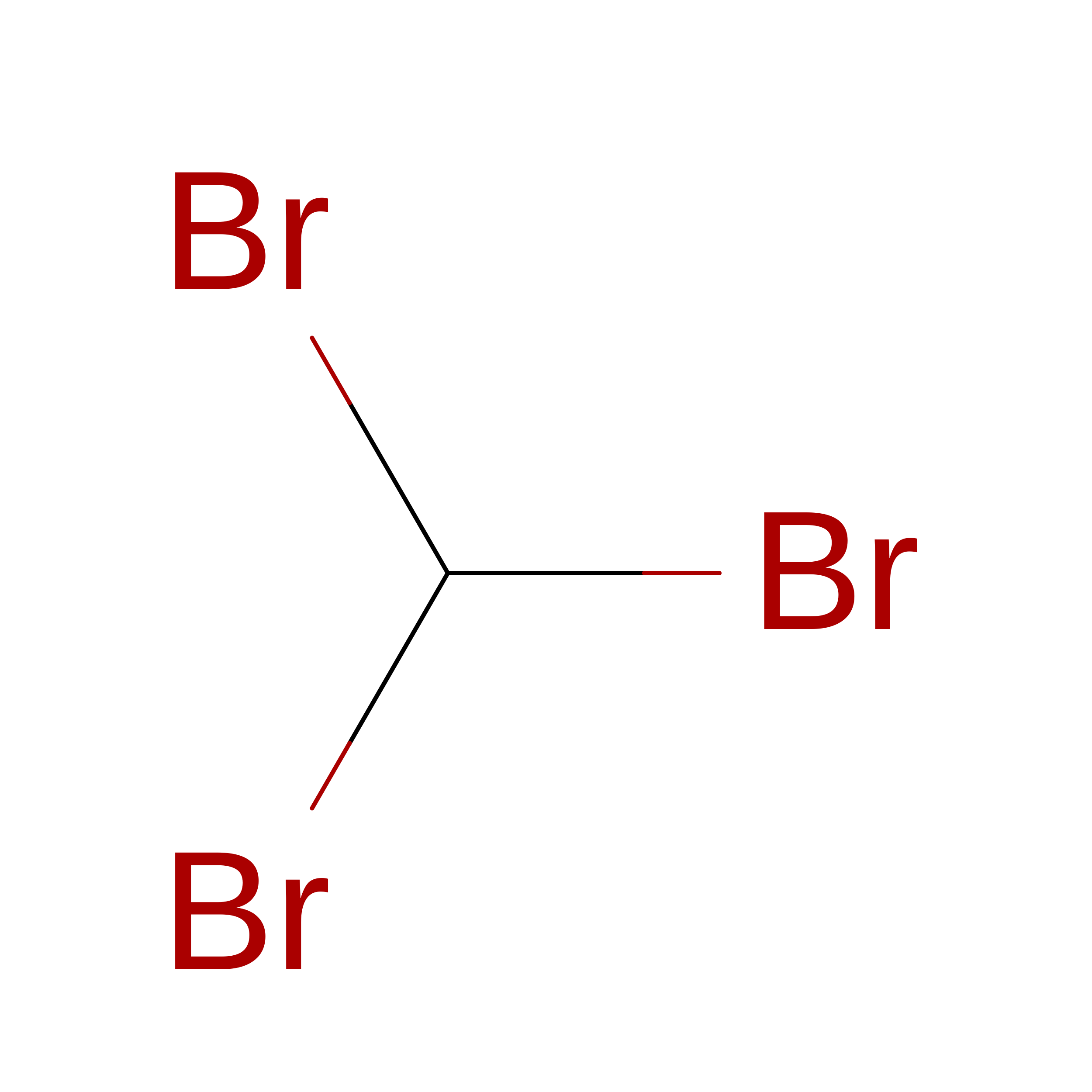
TRIBROMOMETHANE
C H Br3
DIKBFYAXUHHXCS-UHFFFAOYSA-N

Experimental Data & Validation

Experimental Data

  • Method: X-RAY DIFFRACTION
  • Resolution: 3.65 Å
  • R-Value Free:  0.264 (Depositor), 0.267 (DCC) 
  • R-Value Work:  0.229 (Depositor), 0.234 (DCC) 
  • R-Value Observed: 0.231 (Depositor) 
Space Group: P 1 21 1
Unit Cell:
Length ( Å )Angle ( ˚ )
a = 105.109α = 90
b = 266.248β = 109.78
c = 110.746γ = 90
Software Package:
Software NamePurpose
PHENIXrefinement

Structure Validation

View Full Validation Report


Entry History 

Revision History  (Full details and data files)

  • Version 1.0: 2013-02-06
    Type: Initial release
  • Version 1.1: 2013-04-03
    Changes: Database references
  • Version 1.2: 2014-09-17
    Changes: Atomic model, Derived calculations, Non-polymer description, Other
  • Version 1.3: 2024-05-08
    Changes: Data collection, Database references, Derived calculations, Other