3ZHV | pdb_00003zhv

Crystal structure of the SucA domain of Mycobacterium smegmatis KGD, post-decarboxylation intermediate from pyruvate (2-hydroxyethyl-ThDP)


Experimental Data Snapshot

  • Method: X-RAY DIFFRACTION
  • Resolution: 2.30 Å
  • R-Value Free: 
    0.235 (Depositor), 0.235 (DCC) 
  • R-Value Work: 
    0.203 (Depositor) 
  • R-Value Observed: 
    0.205 (Depositor) 

Starting Model: experimental
View more details

wwPDB Validation 3D Report Full Report

Validation slider image for 3ZHV

Ligand Structure Quality Assessment 


This is version 1.2 of the entry. See complete history

Literature

A Dual Conformation of the Post-Decarboxylation Intermediate is Associated with Distinct Enzyme States in Mycobacterial Alpha-Ketoglutarate Decarboxylase (Kgd).

Wagner, T.Barilone, N.Alzari, P.M.Bellinzoni, M.

(2014) Biochem J 457: 425

  • DOI: https://doi.org/10.1042/BJ20131142
  • Primary Citation Related Structures: 
    3ZHQ, 3ZHR, 3ZHS, 3ZHT, 3ZHU, 3ZHV

  • PubMed Abstract: 

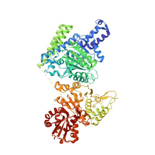
    α-Ketoacid dehydrogenases are large multi-enzyme machineries that orchestrate the oxidative decarboxylation of α-ketoacids with the concomitant production of acyl-CoA and NADH. The first reaction, catalysed by α-ketoacid decarboxylases (E1 enzymes), needs a thiamine diphosphate cofactor and represents the overall rate-limiting step. Although the catalytic cycles of E1 from the pyruvate dehydrogenase (E1p) and branched-chain α-ketoacid dehydrogenase (E1b) complexes have been elucidated, little structural information is available on E1o, the first component of the α-ketoglutarate dehydrogenase complex, despite the central role of this complex at the branching point between the TCA (tricarboxylic acid) cycle and glutamate metabolism. In the present study, we provide structural evidence that MsKGD, the E1o (α-ketoglutarate decarboxylase) from Mycobacterium smegmatis, shows two conformations of the post-decarboxylation intermediate, each one associated with a distinct enzyme state. We also provide an overall picture of the catalytic cycle, reconstructed by either crystallographic snapshots or modelling. The results of the present study show that the conformational change leading the enzyme from the initial (early) to the late state, although not required for decarboxylation, plays an essential role in catalysis and possibly in the regulation of mycobacterial E1o.


  • Organizational Affiliation
    • *Institut Pasteur, Unité de Microbiologie Structurale and CNRS UMR3528, 25 rue du Docteur Roux, 75724 Paris Cedex 15, France.

Macromolecule Content 

  • Total Structure Weight: 390.8 kDa 
  • Atom Count: 26,141 
  • Modeled Residue Count: 3,248 
  • Deposited Residue Count: 3,472 
  • Unique protein chains: 1

Macromolecules

Find similar proteins by:|  3D Structure
Entity ID: 1
MoleculeChains  Sequence LengthOrganismDetailsImage
MULTIFUNCTIONAL 2-OXOGLUTARATE METABOLISM ENZYME
A, B, C, D
868Mycolicibacterium smegmatisMutation(s): 0 
EC: 2.2.1.5 (PDB Primary Data), 4.1.1.71 (PDB Primary Data), 1.2.4.2 (PDB Primary Data), 2.3.1.61 (PDB Primary Data)
UniProt
Find proteins for A0R2B1 (Mycolicibacterium smegmatis (strain ATCC 700084 / mc(2)155))
Explore A0R2B1 
Go to UniProtKB:  A0R2B1
Entity Groups
Sequence Clusters30% Identity50% Identity70% Identity90% Identity95% Identity100% Identity
UniProt GroupA0R2B1
Sequence Annotations
Expand
Reference Sequence

Small Molecules

Ligands 3 Unique
IDChains Name / Formula / InChI Key2D Diagram3D Interactions
TDW

Query on TDW


Download:Ideal Coordinates CCD File
E [auth A],
H [auth B],
K [auth C],
N [auth D]
2-[3-[(4-azanyl-2-methyl-pyrimidin-5-yl)methyl]-4-methyl-2-[(1S)-1-oxidanylethyl]-1,3-thiazol-3-ium-5-yl]ethyl phosphono hydrogen phosphate
C14 H23 N4 O8 P2 S
WHPLAPKWYDKPQH-VIFPVBQESA-N
CA

Query on CA


Download:Ideal Coordinates CCD File
G [auth A],
J [auth B],
M [auth C],
P [auth D]
CALCIUM ION
Ca
BHPQYMZQTOCNFJ-UHFFFAOYSA-N
MG

Query on MG


Download:Ideal Coordinates CCD File
F [auth A],
I [auth B],
L [auth C],
O [auth D]
MAGNESIUM ION
Mg
JLVVSXFLKOJNIY-UHFFFAOYSA-N

Experimental Data & Validation

Experimental Data

  • Method: X-RAY DIFFRACTION
  • Resolution: 2.30 Å
  • R-Value Free:  0.235 (Depositor), 0.235 (DCC) 
  • R-Value Work:  0.203 (Depositor) 
  • R-Value Observed: 0.205 (Depositor) 
Space Group: P 1
Unit Cell:
Length ( Å )Angle ( ˚ )
a = 80.464α = 99.68
b = 83.702β = 98.87
c = 160.333γ = 100.63
Software Package:
Software NamePurpose
BUSTERrefinement
XDSdata reduction
SCALAdata scaling
MOLREPphasing

Structure Validation

View Full Validation Report


Ligand Structure Quality Assessment 


Entry History 

Deposition Data

Revision History  (Full details and data files)

  • Version 1.0: 2013-11-13
    Type: Initial release
  • Version 1.1: 2014-01-29
    Changes: Database references
  • Version 1.2: 2023-12-20
    Changes: Data collection, Database references, Derived calculations, Other, Refinement description