


Crystal Structures of Penicillin-Binding Protein 4 from Listeria Monocytogenes
Jeong, J.H., Kim, Y.G.To be published.
Experimental Data Snapshot
Entity ID: 1 | |||||
|---|---|---|---|---|---|
| Molecule | Chains | Sequence Length | Organism | Details | Image |
| PENICILLIN-BINDING PROTEIN 4 | 47 | Listeria monocytogenes | Mutation(s): 0 | 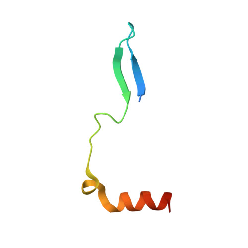 | |
UniProt | |||||
Entity Groups | |||||
| Sequence Clusters | 30% Identity50% Identity70% Identity90% Identity95% Identity100% Identity | ||||
| UniProt Group | Q8Y547 | ||||
Sequence AnnotationsExpand | |||||
Reference Sequence | |||||
Entity ID: 2 | |||||
|---|---|---|---|---|---|
| Molecule | Chains | Sequence Length | Organism | Details | Image |
| PENICILLIN-BINDING PROTEIN 4 | 537 | Listeria monocytogenes | Mutation(s): 0 | 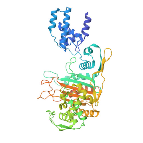 | |
UniProt | |||||
Entity Groups | |||||
| Sequence Clusters | 30% Identity50% Identity70% Identity90% Identity95% Identity100% Identity | ||||
| UniProt Group | Q8Y547 | ||||
Sequence AnnotationsExpand | |||||
Reference Sequence | |||||
| Ligands 2 Unique | |||||
|---|---|---|---|---|---|
| ID | Chains | Name / Formula / InChI Key | 2D Diagram | 3D Interactions | |
| DXF Download:Ideal Coordinates CCD File | C [auth A] | (2R)-5-[(carbamoyloxy)methyl]-2-[(1R)-1-{[(2Z)-2-(furan-2-yl)-2-(methoxyimino)acetyl]amino}-2-oxoethyl]-3,6-dihydro-2H-1,3-thiazine-4-carboxylic acid C16 H18 N4 O8 S RUUDYXYLGTZCPN-VRVPSKPHSA-N |  | ||
| GOL Download:Ideal Coordinates CCD File | D [auth B], E [auth B], F [auth B], G [auth B] | GLYCEROL C3 H8 O3 PEDCQBHIVMGVHV-UHFFFAOYSA-N |  | ||
| Length ( Å ) | Angle ( ˚ ) |
|---|---|
| a = 50.57 | α = 90 |
| b = 78.157 | β = 90 |
| c = 128.914 | γ = 90 |
| Software Name | Purpose |
|---|---|
| PHENIX | refinement |
| HKL-2000 | data reduction |
| SCALEPACK | data scaling |
| PHASER | phasing |


