3ZF8 | pdb_00003zf8

Crystal structure of Saccharomyces cerevisiae Mnn9 in complex with GDP and Mn.


Experimental Data Snapshot

  • Method: X-RAY DIFFRACTION
  • Resolution: 1.98 Å
  • R-Value Free: 
    0.239 (Depositor), 0.242 (DCC) 
  • R-Value Work: 
    0.190 (Depositor), 0.190 (DCC) 
  • R-Value Observed: 
    0.191 (Depositor) 

wwPDB Validation 3D Report Full Report

Validation slider image for 3ZF8

Ligand Structure Quality Assessment 


This is version 1.1 of the entry. See complete history

Literature

Yeast Mnn9 is Both a Priming Glycosyltransferase and an Allosteric Activator of Mannan Biosynthesis.

Striebeck, A.Robinson, D.A.Schuettelkopf, A.W.Van Aalten, D.M.F.

(2013) Open Biol 3: 30022

  • DOI: https://doi.org/10.1098/rsob.130022
  • Primary Citation Related Structures: 
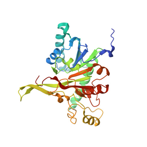
    3ZF8

  • PubMed Abstract: 

    The fungal cell possesses an essential carbohydrate cell wall. The outer layer, mannan, is formed by mannoproteins carrying highly mannosylated O- and N-linked glycans. Yeast mannan biosynthesis is initiated by a Golgi-located complex (M-Pol I) of two GT-62 mannosyltransferases, Mnn9p and Van1p, that are conserved in fungal pathogens. Saccharomyces cerevisiae and Candida albicans mnn9 knockouts show an aberrant cell wall and increased antibiotic sensitivity, suggesting the enzyme is a potential drug target. Here, we present the structure of ScMnn9 in complex with GDP and Mn(2+), defining the fold and catalytic machinery of the GT-62 family. Compared with distantly related GT-78/GT-15 enzymes, ScMnn9 carries an unusual extension. Using a novel enzyme assay and site-directed mutagenesis, we identify conserved amino acids essential for ScMnn9 'priming' α-1,6-mannosyltransferase activity. Strikingly, both the presence of the ScMnn9 protein and its product, but not ScMnn9 catalytic activity, are required to activate subsequent ScVan1 processive α-1,6-mannosyltransferase activity in the M-Pol I complex. These results reveal the molecular basis of mannan synthesis and will aid development of inhibitors targeting this process.


  • Organizational Affiliation
    • Division of Molecular Microbiology, University of Dundee, Dundee DD1 5EH, UK.

Macromolecule Content 

  • Total Structure Weight: 35.54 kDa 
  • Atom Count: 2,506 
  • Modeled Residue Count: 288 
  • Deposited Residue Count: 305 
  • Unique protein chains: 1

Macromolecules

Find similar proteins by:|  3D Structure
Entity ID: 1
MoleculeChains  Sequence LengthOrganismDetailsImage
MANNAN POLYMERASE COMPLEXES SUBUNIT MNN9305Saccharomyces cerevisiae S288CMutation(s): 1 
UniProt
Find proteins for P39107 (Saccharomyces cerevisiae (strain ATCC 204508 / S288c))
Explore P39107 
Go to UniProtKB:  P39107
Entity Groups
Sequence Clusters30% Identity50% Identity70% Identity90% Identity95% Identity100% Identity
UniProt GroupP39107
Sequence Annotations
Expand
Reference Sequence

Small Molecules

Experimental Data & Validation

Experimental Data

  • Method: X-RAY DIFFRACTION
  • Resolution: 1.98 Å
  • R-Value Free:  0.239 (Depositor), 0.242 (DCC) 
  • R-Value Work:  0.190 (Depositor), 0.190 (DCC) 
  • R-Value Observed: 0.191 (Depositor) 
Space Group: P 65 2 2
Unit Cell:
Length ( Å )Angle ( ˚ )
a = 57.09α = 90
b = 57.09β = 90
c = 330.91γ = 120
Software Package:
Software NamePurpose
REFMACrefinement

Structure Validation

View Full Validation Report


Ligand Structure Quality Assessment 


Entry History 

Deposition Data

Revision History  (Full details and data files)

  • Version 1.0: 2013-09-25
    Type: Initial release
  • Version 1.1: 2024-05-08
    Changes: Data collection, Database references, Derived calculations, Other