Structural Insights Into the Intrinsic Self-Assembly of Par-3 N-Terminal Domain.
Zhang, Y., Wang, W., Chen, J., Zhang, K., Gao, F., Gao, B., Zhang, S., Dong, M., Besenbacher, F., Gong, W., Zhang, M., Sun, F., Feng, W.(2013) Structure 21: 997
- PubMed: 23643951 Search on PubMed
- DOI: https://doi.org/10.1016/j.str.2013.04.004
- Primary Citation Related Structures:
3ZEE, 4I6P - PubMed Abstract:
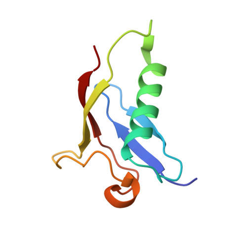
Par-3, the central organizer of the Par-3/Par-6/atypical protein kinase C complex, is a multimodular scaffold protein that is essential for cell polarity establishment and maintenance. The N-terminal domain (NTD) of Par-3 is capable of self-association to form filament-like structures, although the underlying mechanism is poorly understood. Here, we determined the crystal structure of Par-3 NTD and solved the filament structure by cryoelectron microscopy. We found that an intrinsic "front-to-back" interaction mode is important for Par-3 NTD self-association and that both the lateral and longitudinal packing within the filament are mediated by electrostatic interactions. Disruptions of the lateral or longitudinal packing significantly impaired Par-3 NTD self-association and thereby impacted the Par-3-mediated epithelial polarization. We finally demonstrated that a Par-3 NTD-like domain from histidine ammonia-lyase also harbors a similar self-association capacity. This work unequivocally provides the structural basis for Par-3 NTD self-association and characterizes one type of protein domain that can self-assemble via electrostatic interactions.
- National Laboratory of Biomacromolecules, Institute of Biophysics, Chinese Academy of Sciences, 15 Datun Road, Beijing 100101, China.
Organizational Affiliation: