3ZDR | pdb_00003zdr

Structure of the Alcohol dehydrogenase (ADH) domain of a bifunctional ADHE dehydrogenase from Geobacillus thermoglucosidasius NCIMB 11955


Experimental Data Snapshot

  • Method: X-RAY DIFFRACTION
  • Resolution: 2.50 Å
  • R-Value Free: 
    0.240 (Depositor), 0.242 (DCC) 
  • R-Value Work: 
    0.174 (Depositor), 0.181 (DCC) 
  • R-Value Observed: 
    0.177 (Depositor) 

Starting Model: experimental
View more details

wwPDB Validation 3D Report Full Report

Validation slider image for 3ZDR

This is version 2.1 of the entry. See complete history

Literature

Structure of a Bifunctional Alcohol Dehydrogenase Involved in Bioethanol Generation in Geobacillus Thermoglucosidasius

Extance, J.Crennell, S.J.Eley, K.Cripps, R.Hough, D.W.Danson, M.J.

(2013) Acta Crystallogr D Biol Crystallogr 69: 2104

  • DOI: https://doi.org/10.1107/S0907444913020349
  • Primary Citation Related Structures: 
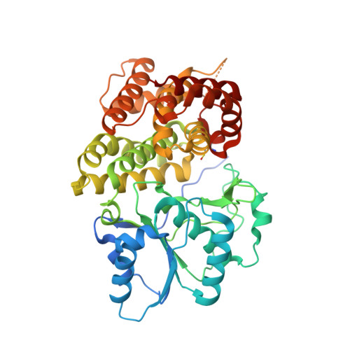
    3ZDR

  • PubMed Abstract: 

    Bifunctional alcohol/aldehyde dehydrogenase (ADHE) enzymes are found within many fermentative microorganisms. They catalyse the conversion of an acyl-coenzyme A to an alcohol via an aldehyde intermediate; this is coupled to the oxidation of two NADH molecules to maintain the NAD(+) pool during fermentative metabolism. The structure of the alcohol dehydrogenase (ADH) domain of an ADHE protein from the ethanol-producing thermophile Geobacillus thermoglucosidasius has been determined to 2.5 Å resolution. This is the first structure to be reported for such a domain. In silico modelling has been carried out to generate a homology model of the aldehyde dehydrogenase domain, and this was subsequently docked with the ADH-domain structure to model the structure of the complete ADHE protein. This model suggests, for the first time, a structural mechanism for the formation of the large multimeric assemblies or `spirosomes' that are observed for this ADHE protein and which have previously been reported for ADHEs from other organisms.


  • Organizational Affiliation
    • Centre for Extremophile Research, Department of Biology and Biochemistry, University of Bath, Bath BA2 7AY, England.

Macromolecule Content 

  • Total Structure Weight: 48.95 kDa 
  • Atom Count: 3,237 
  • Modeled Residue Count: 403 
  • Deposited Residue Count: 433 
  • Unique protein chains: 1

Macromolecules

Find similar proteins by:|  3D Structure
Entity ID: 1
MoleculeChains  Sequence LengthOrganismDetailsImage
ALCOHOL DEHYDROGENASE DOMAIN OF THE BIFUNCTIONAL ACETALDEHYDE DEHYDROGENASE433Parageobacillus thermoglucosidasiusMutation(s): 0 
EC: 1.2.1.10 (PDB Primary Data), 1.1.1.1 (PDB Primary Data)

Experimental Data & Validation

Experimental Data

  • Method: X-RAY DIFFRACTION
  • Resolution: 2.50 Å
  • R-Value Free:  0.240 (Depositor), 0.242 (DCC) 
  • R-Value Work:  0.174 (Depositor), 0.181 (DCC) 
  • R-Value Observed: 0.177 (Depositor) 
Space Group: P 21 21 2
Unit Cell:
Length ( Å )Angle ( ˚ )
a = 73.721α = 90
b = 96.588β = 90
c = 58.2γ = 90
Software Package:
Software NamePurpose
PHENIXrefinement
HKL-2000data reduction
SCALEPACKdata scaling
BALBESphasing

Structure Validation

View Full Validation Report


Entry History 

Deposition Data

Revision History  (Full details and data files)

  • Version 1.0: 2013-10-02
    Type: Initial release
  • Version 1.1: 2013-10-16
    Changes: Database references, Structure summary
  • Version 2.0: 2023-12-20
    Changes: Atomic model, Data collection, Database references, Derived calculations, Other, Refinement description
  • Version 2.1: 2024-11-20
    Changes: Structure summary