3X2P | pdb_00003x2p

Neutron and X-ray joint refined structure of PcCel45A with cellopentaose at 298K.


Experimental Data Snapshot

  • Method: NEUTRON DIFFRACTION
  • Resolution: 1.52 Å
  • R-Value Free: 
    0.260 (Depositor) 
  • R-Value Work: 
    0.218 (Depositor) 
  • R-Value Observed: 
    0.219 (Depositor) 

  • Method: X-RAY DIFFRACTION
  • Resolution: 0.99 Å
  • R-Value Free: 
    0.142 (Depositor), 0.143 (DCC) 
  • R-Value Work: 
    0.134 (Depositor), 0.134 (DCC) 
  • R-Value Observed: 
    0.134 (Depositor) 

wwPDB Validation 3D Report Full Report

Validation slider image for 3X2P

This is version 2.2 of the entry. See complete history

Literature

"Newton's cradle" proton relay with amide-imidic acid tautomerization in inverting cellulase visualized by neutron crystallography.

Nakamura, A.Ishida, T.Kusaka, K.Yamada, T.Fushinobu, S.Tanaka, I.Kaneko, S.Ohta, K.Tanaka, H.Inaka, K.Higuchi, Y.Niimura, N.Samejima, M.Igarashi, K.

(2015) Sci Adv 1: e1500263-e1500263

  • DOI: https://doi.org/10.1126/sciadv.1500263
  • Primary Citation Related Structures: 
    3X2G, 3X2H, 3X2I, 3X2J, 3X2K, 3X2L, 3X2M, 3X2N, 3X2O, 3X2P, 4ZM7

  • PubMed Abstract: 

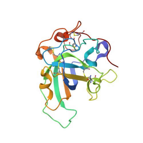
    Hydrolysis of carbohydrates is a major bioreaction in nature, catalyzed by glycoside hydrolases (GHs). We used neutron diffraction and high-resolution x-ray diffraction analyses to investigate the hydrogen bond network in inverting cellulase PcCel45A, which is an endoglucanase belonging to subfamily C of GH family 45, isolated from the basidiomycete Phanerochaete chrysosporium. Examination of the enzyme and enzyme-ligand structures indicates a key role of multiple tautomerizations of asparagine residues and peptide bonds, which are finally connected to the other catalytic residue via typical side-chain hydrogen bonds, in forming the "Newton's cradle"-like proton relay pathway of the catalytic cycle. Amide-imidic acid tautomerization of asparagine has not been taken into account in recent molecular dynamics simulations of not only cellulases but also general enzyme catalysis, and it may be necessary to reconsider our interpretation of many enzymatic reactions.


  • Organizational Affiliation
    • Department of Biomaterials Sciences, Graduate School of Agricultural and Life Sciences, The University of Tokyo, Tokyo 113-8657, Japan.

Macromolecule Content 

  • Total Structure Weight: 19.01 kDa 
  • Atom Count: 1,589 
  • Modeled Residue Count: 180 
  • Deposited Residue Count: 180 
  • Unique protein chains: 1

Macromolecules

Find similar proteins by:|  3D Structure
Entity ID: 1
MoleculeChains  Sequence LengthOrganismDetailsImage
Endoglucanase V-like protein180Phanerodontia chrysosporiumMutation(s): 0 
Gene Names: egvPcCel45A
UniProt
Find proteins for B3Y002 (Phanerodontia chrysosporium)
Explore B3Y002 
Go to UniProtKB:  B3Y002
Entity Groups
Sequence Clusters30% Identity50% Identity70% Identity90% Identity95% Identity100% Identity
UniProt GroupB3Y002
Sequence Annotations
Expand
Reference Sequence

Oligosaccharides

Help  
Entity ID: 2
MoleculeChains Length2D Diagram GlycosylationD Interactions
beta-D-glucopyranose-(1-4)-beta-D-glucopyranose-(1-4)-beta-D-glucopyranose-(1-4)-beta-D-glucopyranose-(1-4)-beta-D-glucopyranose
B
5N/A
Glycosylation Resources
GlyTouCan: G91683DU
GlyCosmos: G91683DU

Small Molecules

Modified Residues  1 Unique
IDChains TypeFormula2D DiagramParent
41Q
Query on 41Q
A
L-PEPTIDE LINKINGC4 H8 N2 O3ASN

Biologically Interesting Molecules (External Reference) 

1 Unique

Experimental Data & Validation

Experimental Data

  • Method: NEUTRON DIFFRACTION
  • Resolution: 1.52 Å
  • R-Value Free:  0.260 (Depositor) 
  • R-Value Work:  0.218 (Depositor) 
  • R-Value Observed: 0.219 (Depositor) 
  • Method: X-RAY DIFFRACTION
  • Resolution: 0.99 Å
  • R-Value Free:  0.142 (Depositor), 0.143 (DCC) 
  • R-Value Work:  0.134 (Depositor), 0.134 (DCC) 
  • R-Value Observed: 0.134 (Depositor) 
Space Group: P 21 21 21
Unit Cell:
Length ( Å )Angle ( ˚ )
a = 46.478α = 90
b = 58.635β = 90
c = 64.744γ = 90
Software Package:
Software NamePurpose
HKL-2000data collection
PHASERphasing
PHENIXrefinement
STARGazerdata reduction
STARGazerdata scaling

Structure Validation

View Full Validation Report


Entry History 

Deposition Data

Revision History  (Full details and data files)

  • Version 1.0: 2015-10-14
    Type: Initial release
  • Version 1.1: 2015-11-04
    Changes: Other
  • Version 1.2: 2018-03-07
    Changes: Data collection
  • Version 1.3: 2018-05-16
    Changes: Data collection
  • Version 1.4: 2019-12-18
    Changes: Database references, Derived calculations
  • Version 2.0: 2020-07-29
    Type: Remediation
    Reason: Carbohydrate remediation
    Changes: Atomic model, Data collection, Derived calculations, Non-polymer description, Structure summary
  • Version 2.1: 2025-03-26
    Changes: Data collection, Database references, Refinement description, Structure summary
  • Version 2.2: 2025-10-29
    Changes: Data collection