3X2F | pdb_00003x2f

A Thermophilic S-Adenosylhomocysteine Hydrolase


Experimental Data Snapshot

  • Method: X-RAY DIFFRACTION
  • Resolution: 2.04 Å
  • R-Value Free: 
    0.230 (Depositor), 0.230 (DCC) 
  • R-Value Work: 
    0.188 (Depositor), 0.188 (DCC) 

Starting Model: experimental
View more details

wwPDB Validation 3D Report Full Report

Validation slider image for 3X2F

Ligand Structure Quality Assessment 


This is version 1.2 of the entry. See complete history

Literature

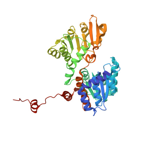
Crystal structures of S-adenosylhomocysteine hydrolase from the thermophilic bacterium Thermotoga maritima.

Zheng, Y.Chen, C.C.Ko, T.P.Xiao, X.Yang, Y.Huang, C.H.Qian, G.Shao, W.Guo, R.T.

(2015) J Struct Biol 190: 135-142

  • DOI: https://doi.org/10.1016/j.jsb.2015.03.002
  • Primary Citation Related Structures: 
    3X2E, 3X2F

  • PubMed Abstract: 

    S-adenosylhomocysteine (SAH) hydrolase catalyzes the reversible hydrolysis of SAH into adenosine and homocysteine by using NAD(+) as a cofactor. The enzyme from Thermotoga maritima (tmSAHH) has great potentials in industrial applications because of its hyperthermophilic properties. Here, two crystal structures of tmSAHH in complex with NAD(+) show both open and closed conformations despite the absence of bound substrate. Each subunit of the tetrameric enzyme is composed of three domains, namely the catalytic domain, the NAD(+)-binding domain and the C-terminal domain. The NAD(+) binding mode is clearly observed and a substrate analogue can also be modeled into the active site, where two cysteine residues in mesophilic enzymes are replaced by serine and threonine in tmSAHH. Notably, the C-terminal domain of tmSAHH lacks the second loop region of mesophilic SAHH, which is important in NAD(+) binding, and thus exposes the bound cofactor to the solvent. The difference explains the higher NAD(+) requirement of tmSAHH because of the reduced affinity. Furthermore, the feature of missing loop is consistently observed in thermophilic bacterial and archaeal SAHHs, and may be related to their thermostability.


  • Organizational Affiliation
    • Industrial Enzymes National Engineering Laboratory, Tianjin Institute of Industrial Biotechnology, Chinese Academy of Sciences, Tianjin 300308, China.

Macromolecule Content 

  • Total Structure Weight: 95.49 kDa 
  • Atom Count: 7,242 
  • Modeled Residue Count: 810 
  • Deposited Residue Count: 822 
  • Unique protein chains: 1

Macromolecules

Find similar proteins by:|  3D Structure
Entity ID: 1
MoleculeChains  Sequence LengthOrganismDetailsImage
Adenosylhomocysteinase
A, B
411Thermotoga maritima MSB8Mutation(s): 0 
Gene Names: ahcYsahhTM_0172
EC: 3.3.1.1 (PDB Primary Data), 3.13.2.1 (UniProt)
UniProt
Find proteins for O51933 (Thermotoga maritima (strain ATCC 43589 / DSM 3109 / JCM 10099 / NBRC 100826 / MSB8))
Explore O51933 
Go to UniProtKB:  O51933
Entity Groups
Sequence Clusters30% Identity50% Identity70% Identity90% Identity95% Identity100% Identity
UniProt GroupO51933
Sequence Annotations
Expand
Reference Sequence

Small Molecules

Ligands 3 Unique
IDChains Name / Formula / InChI Key2D Diagram3D Interactions
NAI

Query on NAI


Download:Ideal Coordinates CCD File
C [auth A],
X [auth B]
1,4-DIHYDRONICOTINAMIDE ADENINE DINUCLEOTIDE
C21 H29 N7 O14 P2
BOPGDPNILDQYTO-NNYOXOHSSA-N
NO3

Query on NO3


Download:Ideal Coordinates CCD File
AA [auth B]
BA [auth B]
CA [auth B]
DA [auth B]
EA [auth B]
AA [auth B],
BA [auth B],
CA [auth B],
DA [auth B],
EA [auth B],
F [auth A],
FA [auth B],
G [auth A],
GA [auth B],
H [auth A],
HA [auth B],
I [auth A],
IA [auth B],
J [auth A],
JA [auth B],
K [auth A],
KA [auth B],
L [auth A],
LA [auth B],
M [auth A],
MA [auth B],
N [auth A],
NA [auth B],
O [auth A],
OA [auth B],
P [auth A],
PA [auth B],
Q [auth A],
QA [auth B],
R [auth A],
RA [auth B],
S [auth A],
SA [auth B],
T [auth A],
TA [auth B],
U [auth A],
UA [auth B],
V [auth A],
W [auth A],
Z [auth B]
NITRATE ION
N O3
NHNBFGGVMKEFGY-UHFFFAOYSA-N
NA

Query on NA


Download:Ideal Coordinates CCD File
D [auth A],
E [auth A],
Y [auth B]
SODIUM ION
Na
FKNQFGJONOIPTF-UHFFFAOYSA-N

Experimental Data & Validation

Experimental Data

  • Method: X-RAY DIFFRACTION
  • Resolution: 2.04 Å
  • R-Value Free:  0.230 (Depositor), 0.230 (DCC) 
  • R-Value Work:  0.188 (Depositor), 0.188 (DCC) 
Space Group: P 31 2 1
Unit Cell:
Length ( Å )Angle ( ˚ )
a = 148.6α = 90
b = 148.6β = 90
c = 84.16γ = 120
Software Package:
Software NamePurpose
HKL-2000data collection
CNSrefinement
HKL-2000data reduction
HKL-2000data scaling
CNSphasing

Structure Validation

View Full Validation Report


Ligand Structure Quality Assessment 


Entry History 

Deposition Data

Revision History  (Full details and data files)

  • Version 1.0: 2015-05-06
    Type: Initial release
  • Version 1.1: 2022-08-24
    Changes: Database references, Derived calculations
  • Version 1.2: 2023-11-08
    Changes: Data collection, Refinement description