3X1J | pdb_00003x1j

Crystal Structure of Phosphopantetheine adenylyltransferase (PPAT/CoaD) with AcCoA from Pseudomonas aeruginosa


Experimental Data Snapshot

  • Method: X-RAY DIFFRACTION
  • Resolution: 2.33 Å
  • R-Value Free: 
    0.278 (Depositor), 0.281 (DCC) 
  • R-Value Work: 
    0.226 (Depositor), 0.229 (DCC) 
  • R-Value Observed: 
    0.228 (Depositor) 

wwPDB Validation 3D Report Full Report

Validation slider image for 3X1J

Ligand Structure Quality Assessment 


This is version 1.3 of the entry. See complete history

Literature

Transition of phosphopantetheine adenylyltransferase from catalytic to allosteric state is characterized by ternary complex formation in Pseudomonas aeruginosa

Chatterjee, R.Mondal, A.Basu, A.Datta, S.

(2016) Biochim Biophys Acta 1864: 773-786

  • DOI: https://doi.org/10.1016/j.bbapap.2016.03.018
  • Primary Citation Related Structures: 
    3X1J, 3X1K, 3X1M, 4RUK

  • PubMed Abstract: 

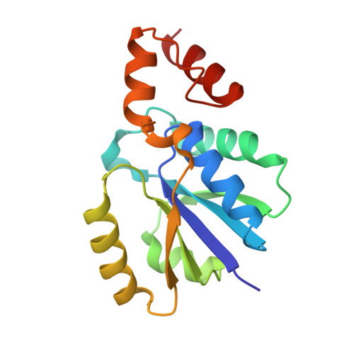
    Phosphopantetheine adenylyltransferase (PPAT) is a rate limiting enzyme which catalyzes the conversion of ATP and pantetheine to dephosphocoenzyme and pyrophosphate. The enzyme is allosteric in nature and regulated by Coenzyme A (CoA) through feedback inhibition. So far, several structures have been solved to decipher the catalytic mechanism of this enzyme. To address catalytic and inhibitory mechanisms of PPAT, structural insights from single crystal X-ray diffraction method were primarily used, followed by biophysical and biochemical analysis. We have solved the structures of PPAT from Pseudomonas aeruginosa with its substrate analogue AMP-PNP and inhibitor CoA. For the first time, a co-crystal structure of PPAT with Acetyl-CoA (AcCoA) was determined. Enzymatic analysis was performed to decipher the catalytic, allosteric and inhibitory mechanisms involved in regulation of PPAT. Binding affinities of PPAT with its substrates and inhibitors were determined by SPR. Previous studies from Escherichia coli and Arabidopsis indicated the inhibitory activity of AcCoA. PPAT-AcCoA structure along with some biochemical methods established AcCoA as an inhibitor to PPAT and illustrated its inhibitory mechanism. Transition from catalytic to allosteric state involves formation of ternary complex. We have studied the structural features of the ternary complex of PPAT along with its product pyrophosphate and inhibitor CoA and validated it with other biophysical and biochemical methods. Extensive analysis of all these 3D structures indicates that changes in side chains R90 and D94 are responsible for transition between catalytic and allosteric inhibitory states. These enzymatic studies provide new insights into the allosteric mechanism of PPAT.


  • Organizational Affiliation
    • Structural Biology and Bioinformatics Division, Council of Scientific and Industrial Research-Indian Institute of Chemical Biology, 4 Raja SC Mullick Road, Jadavpur, Kolkata-700032, West Bengal, India. Electronic address: rakesh.2665675@gmail.com.

Macromolecule Content 

  • Total Structure Weight: 56.92 kDa 
  • Atom Count: 3,983 
  • Modeled Residue Count: 474 
  • Deposited Residue Count: 477 
  • Unique protein chains: 1

Macromolecules

Find similar proteins by:|  3D Structure
Entity ID: 1
MoleculeChains  Sequence LengthOrganismDetailsImage
Phosphopantetheine adenylyltransferase
A, B, C
159Pseudomonas aeruginosa 2192Mutation(s): 0 
Gene Names: coaDPA0363PA2G_04277
EC: 2.7.7.3
UniProt
Find proteins for A0A0X1KGP2 (Pseudomonas aeruginosa 2192)
Explore A0A0X1KGP2 
Go to UniProtKB:  A0A0X1KGP2
Entity Groups
Sequence Clusters30% Identity50% Identity70% Identity90% Identity95% Identity100% Identity
UniProt GroupA0A0X1KGP2
Sequence Annotations
Expand
Reference Sequence

Small Molecules

Ligands 5 Unique
IDChains Name / Formula / InChI Key2D Diagram3D Interactions
ACO

Query on ACO


Download:Ideal Coordinates CCD File
D [auth A],
M [auth B],
Q [auth C]
ACETYL COENZYME *A
C23 H38 N7 O17 P3 S
ZSLZBFCDCINBPY-ZSJPKINUSA-N
PEG

Query on PEG


Download:Ideal Coordinates CCD File
L [auth A]DI(HYDROXYETHYL)ETHER
C4 H10 O3
MTHSVFCYNBDYFN-UHFFFAOYSA-N
PO4

Query on PO4


Download:Ideal Coordinates CCD File
E [auth A],
F [auth A]
PHOSPHATE ION
O4 P
NBIIXXVUZAFLBC-UHFFFAOYSA-K
GOL

Query on GOL


Download:Ideal Coordinates CCD File
G [auth A],
H [auth A]
GLYCEROL
C3 H8 O3
PEDCQBHIVMGVHV-UHFFFAOYSA-N
DMS

Query on DMS


Download:Ideal Coordinates CCD File
I [auth A]
J [auth A]
K [auth A]
N [auth B]
O [auth B]
I [auth A],
J [auth A],
K [auth A],
N [auth B],
O [auth B],
P [auth B],
R [auth C],
S [auth C]
DIMETHYL SULFOXIDE
C2 H6 O S
IAZDPXIOMUYVGZ-UHFFFAOYSA-N

Experimental Data & Validation

Experimental Data

  • Method: X-RAY DIFFRACTION
  • Resolution: 2.33 Å
  • R-Value Free:  0.278 (Depositor), 0.281 (DCC) 
  • R-Value Work:  0.226 (Depositor), 0.229 (DCC) 
  • R-Value Observed: 0.228 (Depositor) 
Space Group: C 2 2 21
Unit Cell:
Length ( Å )Angle ( ˚ )
a = 104.9α = 90
b = 120.7β = 90
c = 90.09γ = 90
Software Package:
Software NamePurpose
MAR345dtbdata collection
PHASERphasing
PHENIXrefinement
MOSFLMdata reduction
SCALAdata scaling

Structure Validation

View Full Validation Report


Ligand Structure Quality Assessment 


Entry History 

Deposition Data

Revision History  (Full details and data files)

  • Version 1.0: 2015-11-25
    Type: Initial release
  • Version 1.1: 2016-04-20
    Changes: Data collection
  • Version 1.2: 2016-05-04
    Changes: Database references
  • Version 1.3: 2024-03-20
    Changes: Data collection, Database references, Derived calculations