3WXM | pdb_00003wxm

Crystal structure of archaeal Pelota and GTP-bound EF1 alpha complex


Experimental Data Snapshot

  • Method: X-RAY DIFFRACTION
  • Resolution: 2.30 Å
  • R-Value Free: 
    0.261 (Depositor), 0.263 (DCC) 
  • R-Value Work: 
    0.198 (Depositor), 0.208 (DCC) 
  • R-Value Observed: 
    0.202 (Depositor) 

wwPDB Validation 3D Report Full Report

Validation slider image for 3WXM

Ligand Structure Quality Assessment 


This is version 1.1 of the entry. See complete history

Literature

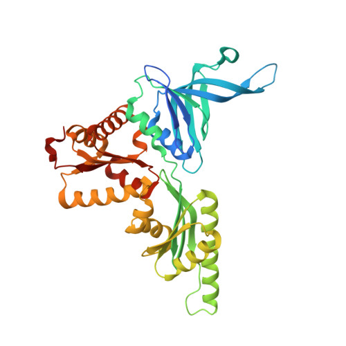
Structural basis for mRNA surveillance by archaeal Pelota and GTP-bound EF1 alpha complex

Kobayashi, K.Kikuno, I.Kuroha, K.Saito, K.Ito, K.Ishitani, R.Inada, T.Nureki, O.

(2010) Proc Natl Acad Sci U S A 107: 17575-17579

  • DOI: https://doi.org/10.1073/pnas.1009598107
  • Primary Citation Related Structures: 
    3WXM

  • PubMed Abstract: 

    No-go decay and nonstop decay are mRNA surveillance pathways that detect translational stalling and degrade the underlying mRNA, allowing the correct translation of the genetic code. In eukaryotes, the protein complex of Pelota (yeast Dom34) and Hbs1 translational GTPase recognizes the stalled ribosome containing the defective mRNA. Recently, we found that archaeal Pelota (aPelota) associates with archaeal elongation factor 1α (aEF1α) to act in the mRNA surveillance pathway, which accounts for the lack of an Hbs1 ortholog in archaea. Here we present the complex structure of aPelota and GTP-bound aEF1α determined at 2.3-Å resolution. The structure reveals how GTP-bound aEF1α recognizes aPelota and how aPelota in turn stabilizes the GTP form of aEF1α. Combined with the functional analysis in yeast, the present results provide structural insights into the molecular interaction between eukaryotic Pelota and Hbs1. Strikingly, the aPelota·aEF1α complex structurally resembles the tRNA·EF-Tu complex bound to the ribosome. Our findings suggest that the molecular mimicry of tRNA in the distorted "A/T state" conformation by Pelota enables the complex to efficiently detect and enter the empty A site of the stalled ribosome.


  • Organizational Affiliation
    • Division of Structure Biology, Department of Basic Medical Sciences, The Institute of Medical Science, University of Tokyo, 4-6-1 Shirokanedai, Minato-ku, Tokyo 108-8639, Japan.

Macromolecule Content 

  • Total Structure Weight: 366.72 kDa 
  • Atom Count: 25,701 
  • Modeled Residue Count: 3,150 
  • Deposited Residue Count: 3,292 
  • Unique protein chains: 2

Macromolecules

Find similar proteins by:|  3D Structure
Entity ID: 1
MoleculeChains  Sequence LengthOrganismDetailsImage
Elongation factor 1-alpha
A, C, E, G
447Aeropyrum pernix K1Mutation(s): 0 
Gene Names: tufAPE_1844
EC: 3.6.5.3
UniProt
Find proteins for Q9YAV0 (Aeropyrum pernix (strain ATCC 700893 / DSM 11879 / JCM 9820 / NBRC 100138 / K1))
Explore Q9YAV0 
Go to UniProtKB:  Q9YAV0
Entity Groups
Sequence Clusters30% Identity50% Identity70% Identity90% Identity95% Identity100% Identity
UniProt GroupQ9YAV0
Sequence Annotations
Expand
Reference Sequence
Find similar proteins by:|  3D Structure
Entity ID: 2
MoleculeChains  Sequence LengthOrganismDetailsImage
Protein pelota homolog
B, D, F, H
376Aeropyrum pernix K1Mutation(s): 0 
Gene Names: pelAAPE_1800.1
EC: 3.1
UniProt
Find proteins for Q9YAZ5 (Aeropyrum pernix (strain ATCC 700893 / DSM 11879 / JCM 9820 / NBRC 100138 / K1))
Explore Q9YAZ5 
Go to UniProtKB:  Q9YAZ5
Entity Groups
Sequence Clusters30% Identity50% Identity70% Identity90% Identity95% Identity100% Identity
UniProt GroupQ9YAZ5
Sequence Annotations
Expand
Reference Sequence

Small Molecules

Ligands 2 Unique
IDChains Name / Formula / InChI Key2D Diagram3D Interactions
GTP

Query on GTP


Download:Ideal Coordinates CCD File
I [auth A],
K [auth C],
M [auth E],
O [auth G]
GUANOSINE-5'-TRIPHOSPHATE
C10 H16 N5 O14 P3
XKMLYUALXHKNFT-UUOKFMHZSA-N
MG

Query on MG


Download:Ideal Coordinates CCD File
J [auth A],
L [auth C],
N [auth E],
P [auth G]
MAGNESIUM ION
Mg
JLVVSXFLKOJNIY-UHFFFAOYSA-N

Experimental Data & Validation

Experimental Data

  • Method: X-RAY DIFFRACTION
  • Resolution: 2.30 Å
  • R-Value Free:  0.261 (Depositor), 0.263 (DCC) 
  • R-Value Work:  0.198 (Depositor), 0.208 (DCC) 
  • R-Value Observed: 0.202 (Depositor) 
Space Group: C 2 2 21
Unit Cell:
Length ( Å )Angle ( ˚ )
a = 148.926α = 90
b = 158.944β = 90
c = 427.236γ = 90
Software Package:
Software NamePurpose
HKL-2000data collection
SHARPphasing
PHENIXrefinement
HKL-2000data reduction
HKL-2000data scaling

Structure Validation

View Full Validation Report


Ligand Structure Quality Assessment 


Entry History 

Deposition Data

Revision History  (Full details and data files)

  • Version 1.0: 2014-09-03
    Type: Initial release
  • Version 1.1: 2024-03-20
    Changes: Data collection, Database references, Derived calculations