3WWQ | pdb_00003wwq

Crystal structure of FAAP20 UBZ domain in complex with Lys63-linked diubiquitin


Experimental Data Snapshot

  • Method: X-RAY DIFFRACTION
  • Resolution: 1.90 Å
  • R-Value Free: 
    0.235 (Depositor), 0.238 (DCC) 
  • R-Value Work: 
    0.208 (Depositor), 0.215 (DCC) 
  • R-Value Observed: 
    0.210 (Depositor) 

Starting Model: experimental
View more details

wwPDB Validation 3D Report Full Report

Validation slider image for 3WWQ

This is version 1.1 of the entry. See complete history

Literature

Structural Basis for Ubiquitin Recognition by Ubiquitin-Binding Zinc Finger of FAAP20

Toma, A.Takahashi, T.S.Sato, Y.Yamagata, A.Goto-Ito, S.Nakada, S.Fukuto, A.Horikoshi, Y.Tashiro, S.Fukai, S.

(2015) PLoS One 10: e0120887-e0120887

  • DOI: https://doi.org/10.1371/journal.pone.0120887
  • Primary Citation Related Structures: 
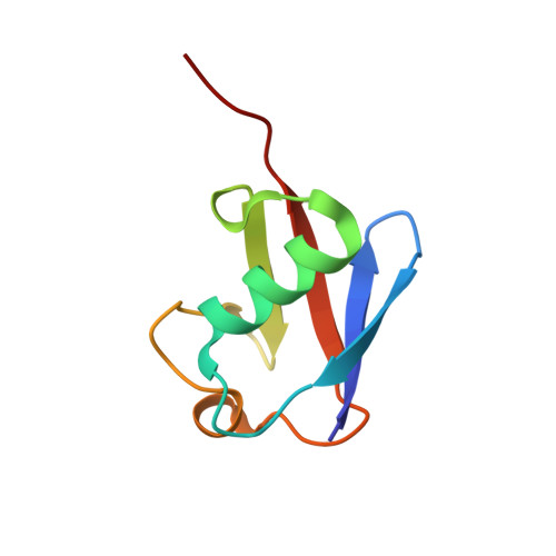
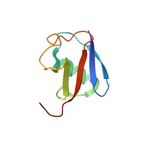
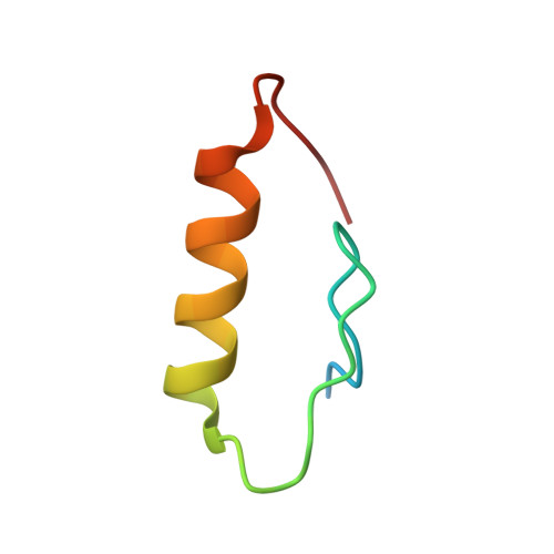
    3WWQ

  • PubMed Abstract: 

    Several ubiquitin-binding zinc fingers (UBZs) have been reported to preferentially bind K63-linked ubiquitin chains. In particular, the UBZ domain of FAAP20 (FAAP20-UBZ), a member of the Fanconi anemia core complex, seems to recognize K63-linked ubiquitin chains, in order to recruit the complex to DNA interstrand crosslinks and mediate DNA repair. By contrast, it is reported that the attachment of a single ubiquitin to Rev1, a translesion DNA polymerase, increases binding of Rev1 to FAAP20. To clarify the specificity of FAAP20-UBZ, we determined the crystal structure of FAAP20-UBZ in complex with K63-linked diubiquitin at 1.9 Å resolution. In this structure, FAAP20-UBZ interacts only with one of the two ubiquitin moieties. Consistently, binding assays using surface plasmon resonance spectrometry showed that FAAP20-UBZ binds ubiquitin and M1-, K48- and K63-linked diubiquitin chains with similar affinities. Residues in the vicinity of Ala168 within the α-helix and the C-terminal Trp180 interact with the canonical Ile44-centered hydrophobic patch of ubiquitin. Asp164 within the α-helix and the C-terminal loop mediate a hydrogen bond network, which reinforces ubiquitin-binding of FAAP20-UBZ. Mutations of the ubiquitin-interacting residues disrupted binding to ubiquitin in vitro and abolished the accumulation of FAAP20 to DNA damage sites in vivo. Finally, structural comparison among FAAP20-UBZ, WRNIP1-UBZ and RAD18-UBZ revealed distinct modes of ubiquitin binding. UBZ family proteins could be divided into at least three classes, according to their ubiquitin-binding modes.


  • Organizational Affiliation
    • Structural Biology Laboratory, Life Science Division, Synchrotron Radiation Research Organization and Institute of Molecular and Cellular Biosciences, The University of Tokyo, Tokyo, 113-0032, Japan; Department of Medical Genome Sciences, Graduate School of Frontier Sciences, The University of Tokyo, Chiba, 277-8501, Japan.

Macromolecule Content 

  • Total Structure Weight: 88.85 kDa 
  • Atom Count: 6,359 
  • Modeled Residue Count: 759 
  • Deposited Residue Count: 788 
  • Unique protein chains: 3

Macromolecules

Find similar proteins by:|  3D Structure
Entity ID: 1
MoleculeChains  Sequence LengthOrganismDetailsImage
Ubiquitin
A, D, G, J
76Mus musculusMutation(s): 1 
Gene Names: Ubc
UniProt & NIH Common Fund Data Resources
Find proteins for P0CG50 (Mus musculus)
Explore P0CG50 
Go to UniProtKB:  P0CG50
IMPC:  MGI:98889
Entity Groups
Sequence Clusters30% Identity50% Identity70% Identity90% Identity95% Identity100% Identity
UniProt GroupP0CG50
Sequence Annotations
Expand
Reference Sequence
Find similar proteins by:|  3D Structure
Entity ID: 2
MoleculeChains  Sequence LengthOrganismDetailsImage
Ubiquitin
B, E, H, K
77Mus musculusMutation(s): 0 
Gene Names: Ubc
UniProt & NIH Common Fund Data Resources
Find proteins for P0CG50 (Mus musculus)
Explore P0CG50 
Go to UniProtKB:  P0CG50
IMPC:  MGI:98889
Entity Groups
Sequence Clusters30% Identity50% Identity70% Identity90% Identity95% Identity100% Identity
UniProt GroupP0CG50
Sequence Annotations
Expand
Reference Sequence
Find similar proteins by:|  3D Structure
Entity ID: 3
MoleculeChains  Sequence LengthOrganismDetailsImage
Fanconi anemia-associated protein of 20 kDa
C, F, I, L
44Homo sapiensMutation(s): 0 
Gene Names: C1orf86FAAP20FP7162
UniProt & NIH Common Fund Data Resources
Find proteins for Q6NZ36 (Homo sapiens)
Explore Q6NZ36 
Go to UniProtKB:  Q6NZ36
PHAROS:  Q6NZ36
GTEx:  ENSG00000162585 
Entity Groups
Sequence Clusters30% Identity50% Identity70% Identity90% Identity95% Identity100% Identity
UniProt GroupQ6NZ36
Sequence Annotations
Expand
Reference Sequence

Experimental Data & Validation

Experimental Data

  • Method: X-RAY DIFFRACTION
  • Resolution: 1.90 Å
  • R-Value Free:  0.235 (Depositor), 0.238 (DCC) 
  • R-Value Work:  0.208 (Depositor), 0.215 (DCC) 
  • R-Value Observed: 0.210 (Depositor) 
Space Group: P 1 21 1
Unit Cell:
Length ( Å )Angle ( ˚ )
a = 59.767α = 90
b = 45.85β = 98.06
c = 172.9γ = 90
Software Package:
Software NamePurpose
MOLREPphasing
REFMACrefinement
HKL-2000data reduction
HKL-2000data scaling

Structure Validation

View Full Validation Report


Entry History 

Deposition Data

Revision History  (Full details and data files)

  • Version 1.0: 2015-05-13
    Type: Initial release
  • Version 1.1: 2023-11-08
    Changes: Data collection, Database references, Derived calculations, Refinement description