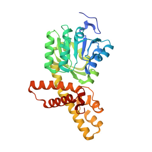
Crystal structure of the NADP+and tartrate-bound complex of L-serine 3-dehydrogenase from the hyperthermophilic archaeon Pyrobaculum calidifontis.
Yoneda, K., Sakuraba, H., Araki, T., Ohshima, T.(2018) Extremophiles 22: 395-405
- PubMed: 29353380
- DOI: https://doi.org/10.1007/s00792-018-1004-0
- Primary Citation of Related Structures:
3W6U, 3W6Z, 3WS7, 5XVH - PubMed Abstract:
A gene encoding L-serine dehydrogenase (L-SerDH) that exhibits extremely low sequence identity to the Agrobacterium tumefaciens L-SerDH was identified in the hyperthermophilic archaeon Pyrobaculum calidifontis. The predicted amino acid sequence showed 36% identity with that of Pseudomonas aeruginosa L-SerDH, suggesting that P. calidifontis L-SerDH is a novel type of L-SerDH, like Ps. aeruginosa L-SerDH. The overexpressed enzyme appears to be the most thermostable L-SerDH described to date, and no loss of activity was observed by incubation for 30 min at temperatures up to 100 °C. The enzyme showed substantial reactivity towards D-serine, in addition to L-serine. Two different crystal structures of P. calidifontis L-SerDH were determined using the Se-MAD and MR method: the structure in complex with NADP + /sulfate ion at 1.18 Å and the structure in complex with NADP + /L-tartrate (substrate analog) at 1.57 Å. The fold of the catalytic domain showed similarity with that of Ps. aeruginosa L-SerDH. However, the active site structure significantly differed between the two enzymes. Based on the structure of the tartrate, L- and D-serine and 3-hydroxypropionate molecules were modeled into the active site and the substrate binding modes were estimated. A structural comparison suggests that the wide cavity at the substrate binding site is likely responsible for the high reactivity of the enzyme toward both L- and D-serine enantiomers. This is the first description of the structure of the novel type of L-SerDH with bound NADP + and substrate analog, and it provides new insight into the substrate binding mechanism of L-SerDH. The results obtained here may be very informative for the creation of L- or D-serine-specific SerDH by protein engineering.
- Department of Bioscience, School of Agriculture, Tokai University, 9-1-1 Toroku, Higashi-ku, Kumamoto, Kumamoto, 862-8652, Japan. kyoneda@agri.u-tokai.ac.jp.
Organizational Affiliation:


