3WQ4 | pdb_00003wq4

Crystal structure of beta-primeverosidase


Experimental Data Snapshot

  • Method: X-RAY DIFFRACTION
  • Resolution: 1.90 Å
  • R-Value Free: 
    0.195 (Depositor), 0.195 (DCC) 
  • R-Value Work: 
    0.159 (Depositor), 0.161 (DCC) 
  • R-Value Observed: 
    0.161 (Depositor) 

Starting Model: experimental
View more details

wwPDB Validation 3D Report Full Report

Validation slider image for 3WQ4

Ligand Structure Quality Assessment 


This is version 2.2 of the entry. See complete history

Literature

Crystal structures of beta-primeverosidase in complex with disaccharide amidine inhibitors.

Saino, H.Shimizu, T.Hiratake, J.Nakatsu, T.Kato, H.Sakata, K.Mizutani, M.

(2014) J Biological Chem 289: 16826-16834

  • DOI: https://doi.org/10.1074/jbc.M114.553271
  • Primary Citation Related Structures: 
    3WQ4, 3WQ5, 3WQ6

  • PubMed Abstract: 

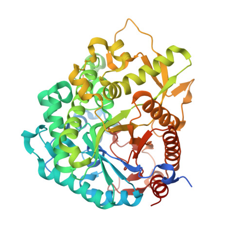
    β-Primeverosidase (PD) is a disaccharide-specific β-glycosidase in tea leaves. This enzyme is involved in aroma formation during the manufacturing process of oolong tea and black tea. PD hydrolyzes β-primeveroside (6-O-β-d-xylopyranosyl-β-d-glucopyranoside) at the β-glycosidic bond of primeverose to aglycone, and releases aromatic alcoholic volatiles of aglycones. PD only accepts primeverose as the glycone substrate, but broadly accepts various aglycones, including 2-phenylethanol, benzyl alcohol, linalool, and geraniol. We determined the crystal structure of PD complexes using highly specific disaccharide amidine inhibitors, N-β-primeverosylamidines, and revealed the architecture of the active site responsible for substrate specificity. We identified three subsites in the active site: subsite -2 specific for 6-O-β-d-xylopyranosyl, subsite -1 well conserved among β-glucosidases and specific for β-d-glucopyranosyl, and wide subsite +1 for hydrophobic aglycone. Glu-470, Ser-473, and Gln-477 act as the specific hydrogen bond donors for 6-O-β-d-xylopyranosyl in subsite -2. On the other hand, subsite +1 was a large hydrophobic cavity that accommodates various aromatic aglycones. Compared with aglycone-specific β-glucosidases of the glycoside hydrolase family 1, PD lacks the Trp crucial for aglycone recognition, and the resultant large cavity accepts aglycone and 6-O-β-d-xylopyranosyl together. PD recognizes the β-primeverosides in subsites -1 and -2 by hydrogen bonds, whereas the large subsite +1 loosely accommodates various aglycones. The glycone-specific activity of PD for broad aglycone substrates results in selective and multiple release of temporally stored alcoholic volatile aglycones of β-primeveroside.


  • Organizational Affiliation
    • From the College of Science and Engineering, Aoyama Gakuin University, Sagamihara-shi, Kanagawa 252-5258, saino@chem.aoyama.ac.jp.

Macromolecule Content 

  • Total Structure Weight: 115.3 kDa 
  • Atom Count: 8,928 
  • Modeled Residue Count: 949 
  • Deposited Residue Count: 1,014 
  • Unique protein chains: 1

Macromolecules

Find similar proteins by:|  3D Structure
Entity ID: 1
MoleculeChains  Sequence LengthOrganismDetailsImage
Beta-primeverosidase
A, B
507Camellia sinensisMutation(s): 0 
EC: 3.2.1.149
UniProt
Find proteins for Q7X9A9 (Camellia sinensis)
Explore Q7X9A9 
Go to UniProtKB:  Q7X9A9
Entity Groups
Sequence Clusters30% Identity50% Identity70% Identity90% Identity95% Identity100% Identity
UniProt GroupQ7X9A9
Glycosylation
Glycosylation Sites: 3
Sequence Annotations
Expand
Reference Sequence

Oligosaccharides

Help  
Entity ID: 2
MoleculeChains Length2D Diagram GlycosylationD Interactions
2-acetamido-2-deoxy-beta-D-glucopyranose-(1-4)-2-acetamido-2-deoxy-beta-D-glucopyranose
C
2N-Glycosylation
Glycosylation Resources
GlyTouCan: G42666HT
GlyCosmos: G42666HT
GlyGen: G42666HT

Experimental Data & Validation

Experimental Data

  • Method: X-RAY DIFFRACTION
  • Resolution: 1.90 Å
  • R-Value Free:  0.195 (Depositor), 0.195 (DCC) 
  • R-Value Work:  0.159 (Depositor), 0.161 (DCC) 
  • R-Value Observed: 0.161 (Depositor) 
Space Group: P 21 21 21
Unit Cell:
Length ( Å )Angle ( ˚ )
a = 59.628α = 90
b = 88.796β = 90
c = 195.17γ = 90
Software Package:
Software NamePurpose
CrystalCleardata collection
MOLREPphasing
PHENIXrefinement
MOSFLMdata reduction
SCALAdata scaling

Structure Validation

View Full Validation Report


Ligand Structure Quality Assessment 


Entry History 

Deposition Data

  • Released Date: 2014-04-23 
  • Deposition Author(s): Saino, H.

Revision History  (Full details and data files)

  • Version 1.0: 2014-04-23
    Type: Initial release
  • Version 1.1: 2015-04-01
    Changes: Database references
  • Version 2.0: 2020-07-29
    Type: Remediation
    Reason: Carbohydrate remediation
    Changes: Atomic model, Data collection, Derived calculations, Structure summary
  • Version 2.1: 2023-11-08
    Changes: Data collection, Database references, Refinement description, Structure summary
  • Version 2.2: 2024-11-20
    Changes: Structure summary