3WNP | pdb_00003wnp

D308A, F268V, D469Y, A513V, and Y515S quintuple mutant of Bacillus circulans T-3040 cycloisomaltooligosaccharide glucanotransferase complexed with isomaltoundecaose


Experimental Data Snapshot

  • Method: X-RAY DIFFRACTION
  • Resolution: 2.80 Å
  • R-Value Free: 
    0.243 (Depositor), 0.244 (DCC) 
  • R-Value Work: 
    0.187 (Depositor), 0.192 (DCC) 
  • R-Value Observed: 
    0.190 (Depositor) 

Starting Model: experimental
View more details

wwPDB Validation 3D Report Full Report

Validation slider image for 3WNP

This is version 2.2 of the entry. See complete history

Literature

Molecular engineering of cycloisomaltooligosaccharide glucanotransferase from Bacillus circulans T-3040: structural determinants for the reaction product size and reactivity.

Suzuki, R.Suzuki, N.Fujimoto, Z.Momma, M.Kimura, K.Kitamura, S.Kimura, A.Funane, K.

(2015) Biochem J 467: 259-270

  • DOI: https://doi.org/10.1042/BJ20140860
  • Primary Citation Related Structures: 
    3WNP

  • PubMed Abstract: 

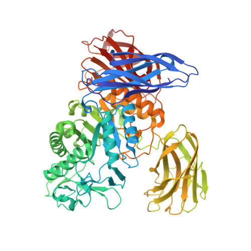
    Cycloisomaltooligosaccharide glucanotransferase (CITase) is a member of glycoside hydrolase family 66 and it produces cycloisomaltooligosaccharides (CIs). Small CIs (CI-7-9) and large CIs (CI-≥10) are designated as oligosaccharide-type CIs (oligo-CIs) and megalosaccharide-type CIs (megalo-CIs) respectively. CITase from Bacillus circulans T-3040 (BcCITase) produces mainly CI-8 with little megalo-CIs. It has two family 35 carbohydrate-binding modules (BcCBM35-1 and BcCBM35-2). BcCBM35-1 is inserted in a catalytic domain of BcCITase and BcCBM35-2 is located at the C-terminal region. Our previous studies suggested that BcCBM35-1 has two substrate-binding sites (B-1 and B-2) [Suzuki et al. (2014) J. Biol. Chem. 289, 12040-12051]. We implemented site-directed mutagenesis of BcCITase to explore the preference for product size on the basis of the 3D structure of BcCITase. Mutational studies provided evidence that B-1 and B-2 contribute to recruiting substrate and maintaining product size respectively. A mutant (mutant-R) with four mutations (F268V, D469Y, A513V and Y515S) produced three times as much megalo-CIs (CI-10-12) and 1.5 times as much total CIs (CI-7-12) as compared with the wild-type (WT) BcCITase. The 3D structure of the substrate-enzyme complex of mutant-R suggested that the modified product size specificity was attributable to the construction of novel substrate-binding sites in the B-2 site of BcCBM35-1 and reactivity was improved by mutation on subsite -3 on the catalytic domain.


  • Organizational Affiliation
    • *Applied Microbiology Division, National Food Research Institute, National Agriculture and Food Research Organization, 2-1-12 Kannondai, Tsukuba 305-8642, Japan.

Macromolecule Content 

  • Total Structure Weight: 161.17 kDa 
  • Atom Count: 11,337 
  • Modeled Residue Count: 1,398 
  • Deposited Residue Count: 1,420 
  • Unique protein chains: 1

Macromolecules

Find similar proteins by:|  3D Structure
Entity ID: 1
MoleculeChains  Sequence LengthOrganismDetailsImage
Cycloisomaltooligosaccharide glucanotransferase
A, B
710Niallia circulansMutation(s): 5 
Gene Names: cit
EC: 2.4.1.248
UniProt
Find proteins for P94286 (Niallia circulans)
Explore P94286 
Go to UniProtKB:  P94286
Entity Groups
Sequence Clusters30% Identity50% Identity70% Identity90% Identity95% Identity100% Identity
UniProt GroupP94286
Sequence Annotations
Expand
Reference Sequence

Oligosaccharides

Help  
Entity ID: 2
MoleculeChains Length2D Diagram GlycosylationD Interactions
alpha-D-glucopyranose-(1-6)-alpha-D-glucopyranose-(1-6)-alpha-D-glucopyranose-(1-6)-beta-D-glucopyranose
C, D
4N/A
Glycosylation Resources
GlyTouCan: G09397NL
GlyCosmos: G09397NL
GlyGen: G09397NL

Small Molecules

Ligands 4 Unique
IDChains Name / Formula / InChI Key2D Diagram3D Interactions
MES

Query on MES


Download:Ideal Coordinates CCD File
M [auth A],
V [auth B]
2-(N-MORPHOLINO)-ETHANESULFONIC ACID
C6 H13 N O4 S
SXGZJKUKBWWHRA-UHFFFAOYSA-N
SO4

Query on SO4


Download:Ideal Coordinates CCD File
G [auth A]
H [auth A]
I [auth A]
J [auth A]
K [auth A]
G [auth A],
H [auth A],
I [auth A],
J [auth A],
K [auth A],
L [auth A],
P [auth B],
Q [auth B],
R [auth B],
S [auth B],
T [auth B],
U [auth B]
SULFATE ION
O4 S
QAOWNCQODCNURD-UHFFFAOYSA-L
CA

Query on CA


Download:Ideal Coordinates CCD File
E [auth A],
N [auth B]
CALCIUM ION
Ca
BHPQYMZQTOCNFJ-UHFFFAOYSA-N
NA

Query on NA


Download:Ideal Coordinates CCD File
F [auth A],
O [auth B]
SODIUM ION
Na
FKNQFGJONOIPTF-UHFFFAOYSA-N

Experimental Data & Validation

Experimental Data

  • Method: X-RAY DIFFRACTION
  • Resolution: 2.80 Å
  • R-Value Free:  0.243 (Depositor), 0.244 (DCC) 
  • R-Value Work:  0.187 (Depositor), 0.192 (DCC) 
  • R-Value Observed: 0.190 (Depositor) 
Space Group: P 21 21 21
Unit Cell:
Length ( Å )Angle ( ˚ )
a = 60.987α = 90
b = 172.813β = 90
c = 172.305γ = 90
Software Package:
Software NamePurpose
SERGUIdata collection
MOLREPphasing
REFMACrefinement
HKL-2000data reduction
SCALEPACKdata scaling

Structure Validation

View Full Validation Report


Entry History 

Deposition Data

Revision History  (Full details and data files)

  • Version 1.0: 2014-02-05
    Type: Initial release
  • Version 1.1: 2015-03-04
    Changes: Database references
  • Version 1.2: 2017-11-22
    Changes: Refinement description
  • Version 2.0: 2020-07-29
    Type: Remediation
    Reason: Carbohydrate remediation
    Changes: Atomic model, Data collection, Database references, Derived calculations, Structure summary
  • Version 2.1: 2022-08-24
    Changes: Database references, Structure summary
  • Version 2.2: 2023-11-08
    Changes: Data collection, Refinement description