3WMQ | pdb_00003wmq

Crystal structure of the complex between SLL-2 and GalNAc.


Experimental Data Snapshot

  • Method: X-RAY DIFFRACTION
  • Resolution: 1.60 Å
  • R-Value Free: 
    0.222 (Depositor), 0.220 (DCC) 
  • R-Value Work: 
    0.212 (Depositor), 0.210 (DCC) 
  • R-Value Observed: 
    0.212 (Depositor) 

Starting Model: experimental
View more details

wwPDB Validation   3D Report Full Report


Ligand Structure Quality Assessment 

Created with Raphaël 2.3.0Worse 01 BetterLigand structure goodness of fit to experimental dataBest fitted A2GClick on this verticalbar to view details

This is version 2.2 of the entry. See complete history


Literature

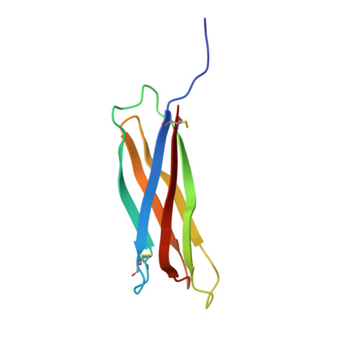
Crystal structure of a symbiosis-related lectin from octocoral.

Kita, A.Jimbo, M.Sakai, R.Morimoto, Y.Miki, K.

(2015) Glycobiology 25: 1016-1023

  • DOI: https://doi.org/10.1093/glycob/cwv033
  • Primary Citation of Related Structures:  
    3WMP, 3WMQ

  • PubMed Abstract: 

    D-Galactose-binding lectin from the octocoral, Sinularia lochmodes (SLL-2), distributes densely on the cell surface of microalgae, Symbiodinium sp., an endosymbiotic dinoflagellate of the coral, and is also shown to be a chemical cue that transforms dinoflagellate into a non-motile (coccoid) symbiotic state. SLL-2 binds with high affinity to the Forssman antigen (N-acetylgalactosamine(GalNAc)α1-3GalNAcβ1-3Galα1-4Galβ1-4Glc-ceramide), and the presence of Forssman antigen-like sugar on the surface of Symbiodinium CS-156 cells was previously confirmed. Here we report the crystal structures of SLL-2 and its GalNAc complex as the first crystal structures of a lectin involved in the symbiosis between coral and dinoflagellate. N-Linked sugar chains and a galactose derivative binding site common to H-type lectins were observed in each monomer of the hexameric SLL-2 crystal structure. In addition, unique sugar-binding site-like regions were identified at the top and bottom of the hexameric SLL-2 structure. These structural features suggest a possible binding mode between SLL-2 and Forssman antigen-like pentasaccharide.


  • Organizational Affiliation

    Research Reactor Institute, Kyoto University, Kumatori, Sennan, Osaka 590-0494, Japan kita@rri.kyoto-u.ac.jp miki@kuchem.kyoto-u.ac.jp.


Macromolecules
Find similar proteins by:  (by identity cutoff)  |  3D Structure
Entity ID: 1
MoleculeChains Sequence LengthOrganismDetailsImage
Galactose-binding lectin94Sclerophytum lochmodesMutation(s): 0 
UniProt
Find proteins for A4CYJ6 (Sinularia lochmodes)
Explore A4CYJ6 
Go to UniProtKB:  A4CYJ6
Entity Groups  
Sequence Clusters30% Identity50% Identity70% Identity90% Identity95% Identity100% Identity
UniProt GroupA4CYJ6
Glycosylation
Glycosylation Sites: 1
Sequence Annotations
Expand
  • Reference Sequence
Oligosaccharides

Help

Entity ID: 2
MoleculeChains Length2D Diagram Glycosylation3D Interactions
2-acetamido-2-deoxy-beta-D-glucopyranose-(1-4)-2-acetamido-2-deoxy-beta-D-glucopyranose
B
2N-Glycosylation
Glycosylation Resources
GlyTouCan:  G42666HT
GlyCosmos:  G42666HT
GlyGen:  G42666HT
Small Molecules
Ligands 4 Unique
IDChains Name / Formula / InChI Key2D Diagram3D Interactions
A2G
Query on A2G

Download Ideal Coordinates CCD File 
C [auth A]2-acetamido-2-deoxy-alpha-D-galactopyranose
C8 H15 N O6
OVRNDRQMDRJTHS-CBQIKETKSA-N
MPD
Query on MPD

Download Ideal Coordinates CCD File 
D [auth A],
E [auth A]
(4S)-2-METHYL-2,4-PENTANEDIOL
C6 H14 O2
SVTBMSDMJJWYQN-YFKPBYRVSA-N
CL
Query on CL

Download Ideal Coordinates CCD File 
F [auth A],
G [auth A],
H [auth A],
I [auth A]
CHLORIDE ION
Cl
VEXZGXHMUGYJMC-UHFFFAOYSA-M
NA
Query on NA

Download Ideal Coordinates CCD File 
J [auth A]SODIUM ION
Na
FKNQFGJONOIPTF-UHFFFAOYSA-N
Experimental Data & Validation

Experimental Data

  • Method: X-RAY DIFFRACTION
  • Resolution: 1.60 Å
  • R-Value Free:  0.222 (Depositor), 0.220 (DCC) 
  • R-Value Work:  0.212 (Depositor), 0.210 (DCC) 
  • R-Value Observed: 0.212 (Depositor) 
Space Group: I 4 3 2
Unit Cell:
Length ( Å )Angle ( ˚ )
a = 154.246α = 90
b = 154.246β = 90
c = 154.246γ = 90
Software Package:
Software NamePurpose
HKL-2000data collection
MOLREPphasing
CNSrefinement
HKL-2000data reduction
SCALEPACKdata scaling

Structure Validation

View Full Validation Report


Ligand Structure Quality Assessment 

Created with Raphaël 2.3.0Worse 01 BetterLigand structure goodness of fit to experimental dataBest fitted A2GClick on this verticalbar to view details

Entry History 

Deposition Data

Revision History  (Full details and data files)

  • Version 1.0: 2015-01-14
    Type: Initial release
  • Version 1.1: 2015-09-23
    Changes: Database references, Structure summary
  • Version 2.0: 2020-07-29
    Type: Remediation
    Reason: Carbohydrate remediation
    Changes: Atomic model, Data collection, Derived calculations, Structure summary
  • Version 2.1: 2023-11-08
    Changes: Data collection, Database references, Refinement description, Structure summary
  • Version 2.2: 2024-11-20
    Changes: Structure summary