3WIZ | pdb_00003wiz

Crystal structure of Bcl-xL in complex with compound 10


Experimental Data Snapshot

  • Method: X-RAY DIFFRACTION
  • Resolution: 2.45 Å
  • R-Value Free: 
    0.238 (Depositor), 0.236 (DCC) 
  • R-Value Work: 
    0.196 (Depositor), 0.196 (DCC) 
  • R-Value Observed: 
    0.198 (Depositor) 

Starting Model: experimental
View more details

wwPDB Validation 3D Report Full Report

Validation slider image for 3WIZ

Ligand Structure Quality Assessment 


This is version 1.3 of the entry. See complete history

Literature

Discovery of potent Mcl-1/Bcl-xL dual inhibitors by using a hybridization strategy based on structural analysis of target proteins.

Tanaka, Y.Aikawa, K.Nishida, G.Homma, M.Sogabe, S.Igaki, S.Hayano, Y.Sameshima, T.Miyahisa, I.Kawamoto, T.Tawada, M.Imai, Y.Inazuka, M.Cho, N.Imaeda, Y.Ishikawa, T.

(2013) J Med Chem 56: 9635-9645

  • DOI: https://doi.org/10.1021/jm401170c
  • Primary Citation Related Structures: 
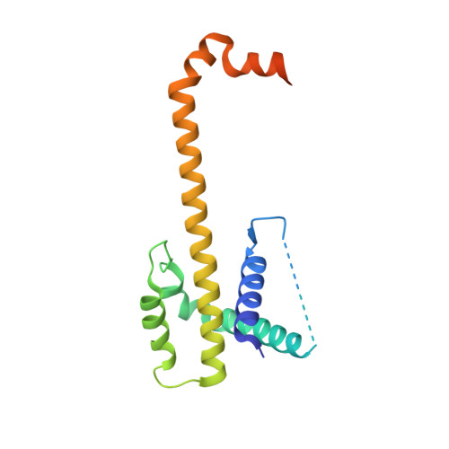
    3WIX, 3WIY, 3WIZ

  • PubMed Abstract: 

    Mcl-1 and Bcl-xL are crucial regulators of apoptosis, therefore dual inhibitors of both proteins could serve as promising new anticancer drugs. To design Mcl-1/Bcl-xL dual inhibitors, we performed structure-guided analyses of the corresponding selective Mcl-1 and Bcl-xL inhibitors. A cocrystal structure of a pyrazolo[1,5-a]pyridine derivative with Mcl-1 protein was successfully determined and revealed the protein-ligand binding mode. The key structure for Bcl-xL inhibition was further confirmed through the substructural analysis of ABT-263, a representative Bcl-xL/Bcl-2/Bcl-w inhibitor developed by Abbott Laboratories. On the basis of the structural data from this analysis, we designed hybrid compounds by tethering the Mcl-1 and Bcl-xL inhibitors together. The results of X-ray crystallographic analysis of hybrid compound 10 in complexes with both Mcl-1 and Bcl-xL demonstrated its binding mode with each protein. Following further optimization, compound 11 showed potent Mcl-1/Bcl-xL dual inhibitory activity (Mcl-1, IC50 = 0.088 μM; and Bcl-xL, IC50 = 0.0037 μM).


  • Organizational Affiliation
    • Pharmaceutical Research Division, Takeda Pharmaceutical Company Limited , 26-1, Muraoka-Higashi 2-chome, Fujisawa, Kanagawa 251-8555, Japan.

Macromolecule Content 

  • Total Structure Weight: 42.73 kDa 
  • Atom Count: 2,453 
  • Modeled Residue Count: 282 
  • Deposited Residue Count: 354 
  • Unique protein chains: 1

Macromolecules

Find similar proteins by:|  3D Structure
Entity ID: 1
MoleculeChains  Sequence LengthOrganismDetailsImage
Bcl-2-like protein 1
A, B
177Homo sapiensMutation(s): 0 
Gene Names: BCL2L1BCL2LBCLX
UniProt & NIH Common Fund Data Resources
Find proteins for Q07817 (Homo sapiens)
Explore Q07817 
Go to UniProtKB:  Q07817
PHAROS:  Q07817
GTEx:  ENSG00000171552 
Entity Groups
Sequence Clusters30% Identity50% Identity70% Identity90% Identity95% Identity100% Identity
UniProt GroupQ07817
Sequence Annotations
Expand
Reference Sequence

Small Molecules

Ligands 2 Unique
IDChains Name / Formula / InChI Key2D Diagram3D Interactions
LC6

Query on LC6


Download:Ideal Coordinates CCD File
C [auth A],
E [auth B]
7-(4-{[(4-{[(2R)-4-(dimethylamino)-1-(phenylsulfanyl)butan-2-yl]amino}-3-nitrophenyl)sulfonyl]carbamoyl}-2-methylphenyl)-3-[3-(naphthalen-1-yloxy)propyl]pyrazolo[1,5-a]pyridine-2-carboxylic acid
C47 H46 N6 O8 S2
BNBDPYUSBJEPGX-UUWRZZSWSA-N
PO4

Query on PO4


Download:Ideal Coordinates CCD File
D [auth A],
F [auth B]
PHOSPHATE ION
O4 P
NBIIXXVUZAFLBC-UHFFFAOYSA-K

Experimental Data & Validation

Experimental Data

  • Method: X-RAY DIFFRACTION
  • Resolution: 2.45 Å
  • R-Value Free:  0.238 (Depositor), 0.236 (DCC) 
  • R-Value Work:  0.196 (Depositor), 0.196 (DCC) 
  • R-Value Observed: 0.198 (Depositor) 
Space Group: I 21 3
Unit Cell:
Length ( Å )Angle ( ˚ )
a = 152.661α = 90
b = 152.661β = 90
c = 152.661γ = 90
Software Package:
Software NamePurpose
MOLREPphasing
REFMACrefinement
HKL-2000data reduction
HKL-2000data scaling

Structure Validation

View Full Validation Report


Ligand Structure Quality Assessment 


Entry History 

Deposition Data

Revision History  (Full details and data files)

  • Version 1.0: 2013-11-27
    Type: Initial release
  • Version 1.1: 2014-03-12
    Changes: Database references
  • Version 1.2: 2017-08-23
    Changes: Refinement description, Source and taxonomy
  • Version 1.3: 2023-11-08
    Changes: Data collection, Database references, Derived calculations, Refinement description