3WFL | pdb_00003wfl

Crtstal structure of glycoside hydrolase family 5 beta-mannanase from Talaromyces trachyspermus


Experimental Data Snapshot

  • Method: X-RAY DIFFRACTION
  • Resolution: 1.60 Å
  • R-Value Free: 
    0.138 (Depositor), 0.161 (DCC) 
  • R-Value Work: 
    0.114 (Depositor) 
  • R-Value Observed: 
    0.115 (Depositor) 

Starting Model: experimental
View more details

wwPDB Validation 3D Report Full Report

Validation slider image for 3WFL

Ligand Structure Quality Assessment 


This is version 1.3 of the entry. See complete history

Literature

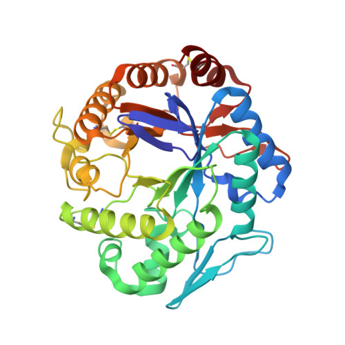
Purification, cloning, functional expression, structure, and characterization of a thermostable beta-mannanase from Talaromyces trachyspermus and its efficiency in production of mannooligosaccharides from coffee wastes

Ichinose, H.Suzuki, K.Yuki, M.Michikawa, M.Sato, H.Kamino, K.Ogasawara, W.Fushinobu, S.Kaneko, S.

To be published.

Macromolecule Content 

  • Total Structure Weight: 38.27 kDa 
  • Atom Count: 3,128 
  • Modeled Residue Count: 341 
  • Deposited Residue Count: 342 
  • Unique protein chains: 1

Macromolecules

Find similar proteins by:|  3D Structure
Entity ID: 1
MoleculeChains  Sequence LengthOrganismDetailsImage
beta-mannanase342Talaromyces trachyspermusMutation(s): 0 
EC: 3.2.1.78
UniProt
Find proteins for A0A087WNH1 (Talaromyces trachyspermus)
Explore A0A087WNH1 
Go to UniProtKB:  A0A087WNH1
Entity Groups
Sequence Clusters30% Identity50% Identity70% Identity90% Identity95% Identity100% Identity
UniProt GroupA0A087WNH1
Glycosylation
Glycosylation Sites: 1
Sequence Annotations
Expand
Reference Sequence

Small Molecules

Ligands 4 Unique
IDChains Name / Formula / InChI Key2D Diagram3D Interactions
NAG

Query on NAG


Download:Ideal Coordinates CCD File
B [auth A]2-acetamido-2-deoxy-beta-D-glucopyranose
C8 H15 N O6
OVRNDRQMDRJTHS-FMDGEEDCSA-N
TRS

Query on TRS


Download:Ideal Coordinates CCD File
C [auth A]2-AMINO-2-HYDROXYMETHYL-PROPANE-1,3-DIOL
C4 H12 N O3
LENZDBCJOHFCAS-UHFFFAOYSA-O
PO4

Query on PO4


Download:Ideal Coordinates CCD File
K [auth A],
L [auth A]
PHOSPHATE ION
O4 P
NBIIXXVUZAFLBC-UHFFFAOYSA-K
GOL

Query on GOL


Download:Ideal Coordinates CCD File
D [auth A]
E [auth A]
F [auth A]
G [auth A]
H [auth A]
D [auth A],
E [auth A],
F [auth A],
G [auth A],
H [auth A],
I [auth A],
J [auth A]
GLYCEROL
C3 H8 O3
PEDCQBHIVMGVHV-UHFFFAOYSA-N

Experimental Data & Validation

Experimental Data

  • Method: X-RAY DIFFRACTION
  • Resolution: 1.60 Å
  • R-Value Free:  0.138 (Depositor), 0.161 (DCC) 
  • R-Value Work:  0.114 (Depositor) 
  • R-Value Observed: 0.115 (Depositor) 
Space Group: P 21 21 21
Unit Cell:
Length ( Å )Angle ( ˚ )
a = 58.434α = 90
b = 75.972β = 90
c = 79.723γ = 90
Software Package:
Software NamePurpose
HKL-2000data collection
MOLREPphasing
REFMACrefinement
HKL-2000data reduction
HKL-2000data scaling

Structure Validation

View Full Validation Report


Ligand Structure Quality Assessment 


Entry History 

Deposition Data

Revision History  (Full details and data files)

  • Version 1.0: 2014-07-23
    Type: Initial release
  • Version 1.1: 2020-07-29
    Type: Remediation
    Reason: Carbohydrate remediation
    Changes: Data collection, Derived calculations, Structure summary
  • Version 1.2: 2023-11-08
    Changes: Data collection, Database references, Refinement description, Structure summary
  • Version 1.3: 2024-10-09
    Changes: Structure summary