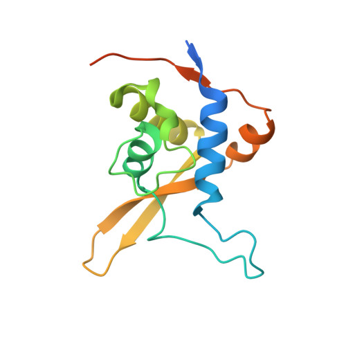
3WE2

Structure of BLM RQC domain bound to a phosphate ion


Experimental Data Snapshot

  • Method: X-RAY DIFFRACTION
  • Resolution: 2.70 Å
  • R-Value Free: 0.271 
  • R-Value Work: 0.237 

wwPDB Validation   3D Report Full Report


This is version 1.2 of the entry. See complete history


Literature

Structure of the RecQ C-terminal Domain of Human Bloom Syndrome Protein

Kim, S.Y.Hakoshima, T.Kitano, K.

(2013) Sci Rep 3: 3294-3294

  • DOI: https://doi.org/10.1038/srep03294
  • Primary Citation of Related Structures:  
    3WE2, 3WE3

  • PubMed Abstract: 

    Bloom syndrome is a rare genetic disorder characterized by genomic instability and cancer predisposition. The disease is caused by mutations of the Bloom syndrome protein (BLM). Here we report the crystal structure of a RecQ C-terminal (RQC) domain from human BLM. The structure reveals three novel features of BLM RQC which distinguish it from the previous structures of the Werner syndrome protein (WRN) and RECQ1. First, BLM RQC lacks an aromatic residue at the tip of the β-wing, a key element of the RecQ-family helicases used for DNA-strand separation. Second, a BLM-specific insertion between the N-terminal helices exhibits a looping-out structure that extends at right angles to the β-wing. Deletion mutagenesis of this insertion interfered with binding to Holliday junction. Third, the C-terminal region of BLM RQC adopts an extended structure running along the domain surface, which may facilitate the spatial positioning of an HRDC domain in the full-length protein.


  • Organizational Affiliation

    Structural Biology Laboratory, Graduate School of Biological Sciences, Nara Institute of Science and Technology, 8916-5 Takayama, Ikoma, Nara 630-0192, Japan.


Macromolecules
Find similar proteins by:  (by identity cutoff)  |  3D Structure
Entity ID: 1
MoleculeChains Sequence LengthOrganismDetailsImage
Bloom syndrome protein
A, B
147Homo sapiensMutation(s): 0 
Gene Names: BLM
EC: 3.6.4.12 (PDB Primary Data), 5.6.2.4 (UniProt)
UniProt & NIH Common Fund Data Resources
Find proteins for P54132 (Homo sapiens)
Explore P54132 
Go to UniProtKB:  P54132
PHAROS:  P54132
GTEx:  ENSG00000197299 
Entity Groups  
Sequence Clusters30% Identity50% Identity70% Identity90% Identity95% Identity100% Identity
UniProt GroupP54132
Sequence Annotations
Expand
  • Reference Sequence
Experimental Data & Validation

Experimental Data

  • Method: X-RAY DIFFRACTION
  • Resolution: 2.70 Å
  • R-Value Free: 0.271 
  • R-Value Work: 0.237 
  • Space Group: P 43 21 2
Unit Cell:
Length ( Å )Angle ( ˚ )
a = 59.218α = 90
b = 59.218β = 90
c = 210.146γ = 90
Software Package:
Software NamePurpose
HKL-2000data collection
SHARPphasing
CNSrefinement
HKL-2000data reduction
HKL-2000data scaling

Structure Validation

View Full Validation Report


Entry History 

Deposition Data

Revision History  (Full details and data files)

  • Version 1.0: 2013-11-13
    Type: Initial release
  • Version 1.1: 2013-12-04
    Changes: Database references, Structure summary
  • Version 1.2: 2024-03-20
    Changes: Data collection, Database references, Derived calculations