3WDZ | pdb_00003wdz

Crystal Structure of Keap1 in Complex with phosphorylated p62


Experimental Data Snapshot

  • Method: X-RAY DIFFRACTION
  • Resolution: 2.60 Å
  • R-Value Free: 
    0.255 (Depositor), 0.254 (DCC) 
  • R-Value Work: 
    0.191 (Depositor), 0.189 (DCC) 
  • R-Value Observed: 
    0.194 (Depositor) 

Starting Model: experimental
View more details

wwPDB Validation 3D Report Full Report

Validation slider image for 3WDZ

This is version 1.3 of the entry. See complete history

Literature

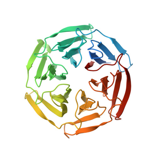
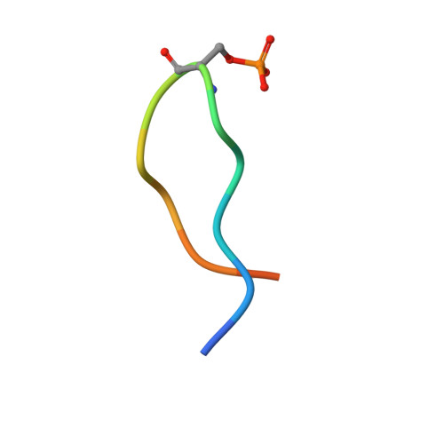
Phosphorylation of p62 activates the Keap1-Nrf2 pathway during selective autophagy.

Ichimura, Y.Waguri, S.Sou, Y.S.Kageyama, S.Hasegawa, J.Ishimura, R.Saito, T.Yang, Y.Kouno, T.Fukutomi, T.Hoshii, T.Hirao, A.Takagi, K.Mizushima, T.Motohashi, H.Lee, M.S.Yoshimori, T.Tanaka, K.Yamamoto, M.Komatsu, M.

(2013) Mol Cell 51: 618-631

  • DOI: https://doi.org/10.1016/j.molcel.2013.08.003
  • Primary Citation Related Structures: 
    3WDZ

  • PubMed Abstract: 

    The Keap1-Nrf2 system and autophagy are both involved in the oxidative-stress response, metabolic pathways, and innate immunity, and dysregulation of these processes is associated with pathogenic processes. However, the interplay between these two pathways remains largely unknown. Here, we show that phosphorylation of the autophagy-adaptor protein p62 markedly increases p62's binding affinity for Keap1, an adaptor of the Cul3-ubiquitin E3 ligase complex responsible for degrading Nrf2. Thus, p62 phosphorylation induces expression of cytoprotective Nrf2 targets. p62 is assembled on selective autophagic cargos such as ubiquitinated organelles and subsequently phosphorylated in an mTORC1-dependent manner, implying coupling of the Keap1-Nrf2 system to autophagy. Furthermore, persistent activation of Nrf2 through accumulation of phosphorylated p62 contributes to the growth of human hepatocellular carcinomas (HCCs). These results demonstrate that selective autophagy and the Keap1-Nrf2 pathway are interdependent, and that inhibitors of the interaction between phosphorylated p62 and Keap1 have potential as therapeutic agents against human HCC.


  • Organizational Affiliation
    • Protein Metabolism Project, Tokyo Metropolitan Institute of Medical Science, Tokyo 156-8506, Japan.

Macromolecule Content 

  • Total Structure Weight: 35.88 kDa 
  • Atom Count: 2,281 
  • Modeled Residue Count: 296 
  • Deposited Residue Count: 325 
  • Unique protein chains: 2

Macromolecules

Find similar proteins by:|  3D Structure
Entity ID: 1
MoleculeChains  Sequence LengthOrganismDetailsImage
Kelch-like ECH-associated protein 1311Mus musculusMutation(s): 0 
Gene Names: Keap1Inrf2Kiaa0132
UniProt
Find proteins for Q9Z2X8 (Mus musculus)
Explore Q9Z2X8 
Go to UniProtKB:  Q9Z2X8
Entity Groups
Sequence Clusters30% Identity50% Identity70% Identity90% Identity95% Identity100% Identity
UniProt GroupQ9Z2X8
Sequence Annotations
Expand
Reference Sequence
Find similar proteins by:  Sequence   |   3D Structure  
Entity ID: 2
MoleculeChains  Sequence LengthOrganismDetailsImage
Peptide from Sequestosome-114Mus musculusMutation(s): 0 
UniProt & NIH Common Fund Data Resources
Find proteins for Q64337 (Mus musculus)
Explore Q64337 
Go to UniProtKB:  Q64337
IMPC:  MGI:107931
Entity Groups
Sequence Clusters30% Identity50% Identity70% Identity90% Identity95% Identity100% Identity
UniProt GroupQ64337
Sequence Annotations
Expand
Reference Sequence

Small Molecules

Modified Residues  1 Unique
IDChains TypeFormula2D DiagramParent
SEP
Query on SEP
B
L-PEPTIDE LINKINGC3 H8 N O6 PSER

Experimental Data & Validation

Experimental Data

  • Method: X-RAY DIFFRACTION
  • Resolution: 2.60 Å
  • R-Value Free:  0.255 (Depositor), 0.254 (DCC) 
  • R-Value Work:  0.191 (Depositor), 0.189 (DCC) 
  • R-Value Observed: 0.194 (Depositor) 
Space Group: P 32 2 1
Unit Cell:
Length ( Å )Angle ( ˚ )
a = 75.149α = 90
b = 75.149β = 90
c = 113.008γ = 120
Software Package:
Software NamePurpose
MOLREPphasing
REFMACrefinement
HKL-2000data reduction
HKL-2000data scaling

Structure Validation

View Full Validation Report


Entry History 

Deposition Data

Revision History  (Full details and data files)

  • Version 1.0: 2013-09-04
    Type: Initial release
  • Version 1.1: 2013-12-25
    Changes: Database references, Source and taxonomy
  • Version 1.2: 2023-11-08
    Changes: Data collection, Database references, Derived calculations, Refinement description
  • Version 1.3: 2024-11-13
    Changes: Structure summary