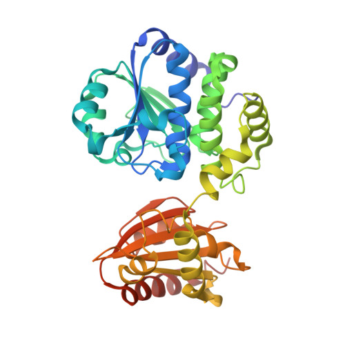
3W5W | pdb_00003w5w

Mn2+-GMP complex of nanoRNase (Nrn) from Bacteroides fragilis


Experimental Data Snapshot

  • Method: X-RAY DIFFRACTION
  • Resolution: 2.95 Å
  • R-Value Free: 
    0.270 (Depositor), 0.210 (DCC) 
  • R-Value Work: 
    0.215 (Depositor), 0.210 (DCC) 

Starting Model: experimental
View more details

wwPDB Validation   3D Report Full Report


Ligand Structure Quality Assessment 

Created with Raphaël 2.3.0Worse 01 BetterLigand structure goodness of fit to experimental dataBest fitted 5GPClick on this verticalbar to view details

This is version 1.2 of the entry. See complete history


Literature

Crystal structure of the ligand-binding form of nanoRNase from Bacteroides fragilis, a member of the DHH/DHHA1 phosphoesterase family of proteins.

Uemura, Y.Nakagawa, N.Wakamatsu, T.Kim, K.Montelione, G.T.Hunt, J.F.Kuramitsu, S.Masui, R.

(2013) FEBS Lett 587: 2669-2674

  • DOI: https://doi.org/10.1016/j.febslet.2013.06.053
  • Primary Citation of Related Structures:  
    3W5W

  • PubMed Abstract: 

    NanoRNase (Nrn) specifically degrades nucleoside 3',5'-bisphosphate and the very short RNA, nanoRNA, during the final step of mRNA degradation. The crystal structure of Nrn in complex with a reaction product GMP was determined. The overall structure consists of two domains that are interconnected by a flexible loop and form a cleft. Two Mn²⁺ ions are coordinated by conserved residues in the DHH motif of the N-terminal domain. GMP binds near the DHHA1 motif region in the C-terminal domain. Our structure enables us to predict the substrate-bound form of Nrn as well as other DHH/DHHA1 phosphoesterase family proteins.


  • Organizational Affiliation

    Graduate School of Frontier Biosciences, Osaka University, Suita, Osaka 565-0871, Japan.


Macromolecules
Find similar proteins by:  (by identity cutoff)  |  3D Structure
Entity ID: 1
MoleculeChains Sequence LengthOrganismDetailsImage
Putative exopolyphosphatase-related protein351Bacteroides fragilis YCH46Mutation(s): 0 
Gene Names: BF3900
UniProt
Find proteins for Q64PD9 (Bacteroides fragilis (strain YCH46))
Explore Q64PD9 
Go to UniProtKB:  Q64PD9
Entity Groups  
Sequence Clusters30% Identity50% Identity70% Identity90% Identity95% Identity100% Identity
UniProt GroupQ64PD9
Sequence Annotations
Expand
  • Reference Sequence
Experimental Data & Validation

Experimental Data

  • Method: X-RAY DIFFRACTION
  • Resolution: 2.95 Å
  • R-Value Free:  0.270 (Depositor), 0.210 (DCC) 
  • R-Value Work:  0.215 (Depositor), 0.210 (DCC) 
Space Group: P 32 2 1
Unit Cell:
Length ( Å )Angle ( ˚ )
a = 90.828α = 90
b = 90.828β = 90
c = 107.878γ = 120
Software Package:
Software NamePurpose
SCALEPACKdata scaling
CNSrefinement
PDB_EXTRACTdata extraction
HKL-2000data reduction
HKL-2000data scaling
MOLREPphasing

Structure Validation

View Full Validation Report


Ligand Structure Quality Assessment 

Created with Raphaël 2.3.0Worse 01 BetterLigand structure goodness of fit to experimental dataBest fitted 5GPClick on this verticalbar to view details

Entry History 

Deposition Data

Revision History  (Full details and data files)

  • Version 1.0: 2013-07-10
    Type: Initial release
  • Version 1.1: 2022-08-24
    Changes: Database references, Derived calculations
  • Version 1.2: 2023-11-08
    Changes: Data collection, Refinement description