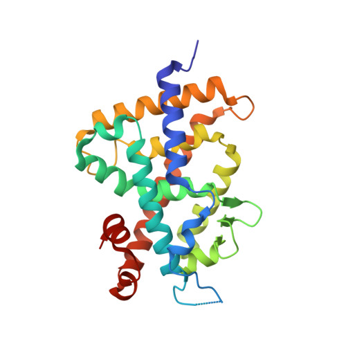
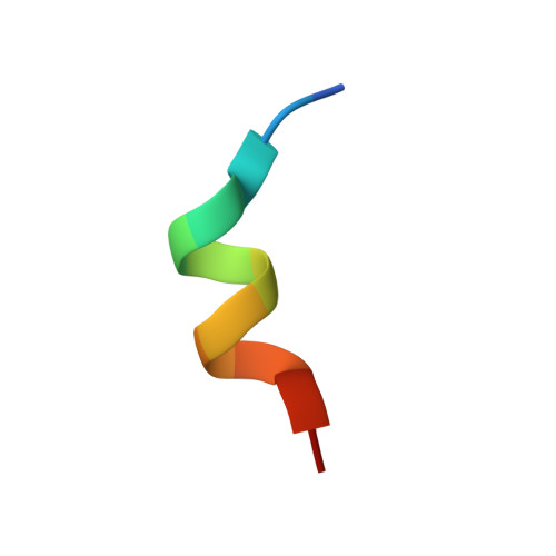
3W0G

Crystal Structure of Rat VDR Ligand Binding Domain in Complex with Novel Nonsecosteroidal Ligands


Experimental Data Snapshot

  • Method: X-RAY DIFFRACTION
  • Resolution: 1.94 Å
  • R-Value Free: 0.258 
  • R-Value Work: 0.214 
  • R-Value Observed: 0.216 

Starting Model: experimental
View more details

wwPDB Validation   3D Report Full Report


Ligand Structure Quality Assessment 


This is version 1.2 of the entry. See complete history


Literature

Structural basis for vitamin D receptor agonism by novel non-secosteroidal ligands.

Asano, L.Ito, I.Kuwabara, N.Waku, T.Yanagisawa, J.Miyachi, H.Shimizu, T.

(2013) FEBS Lett 587: 957-963

  • DOI: https://doi.org/10.1016/j.febslet.2013.02.028
  • Primary Citation of Related Structures:  
    3W0G, 3W0H, 3W0I, 3W0J

  • PubMed Abstract: 

    Non-secosteroidal ligands for vitamin D receptor (VDR) have been developed for the agonist with non-calcemic profiles. Here, we provide the structural mechanism of VDR agonism by novel non-secosteroidal ligands. All ligands had the similar efficacy, while two had the higher potency. Crystallographic analyses revealed that all ligands interacted with helix H10 and the loop between helices H6 and H7 in a similar manner, but also that the two ligands with higher potency had different interaction modes. This study suggests that distinct ligand potency depend upon differences in the formation and rearrangement of hydrogen-bond networks induced by each ligand.


  • Organizational Affiliation

    Graduate School of Pharmaceutical Sciences, University of Tokyo, Hongo, Bunkyo-ku, Tokyo, Japan.


Macromolecules
Find similar proteins by:  (by identity cutoff)  |  3D Structure
Entity ID: 1
MoleculeChains Sequence LengthOrganismDetailsImage
Vitamin D3 receptor253Rattus norvegicusMutation(s): 0 
Gene Names: Nr1i1Vdr
UniProt
Find proteins for P13053 (Rattus norvegicus)
Explore P13053 
Go to UniProtKB:  P13053
Entity Groups  
Sequence Clusters30% Identity50% Identity70% Identity90% Identity95% Identity100% Identity
UniProt GroupP13053
Sequence Annotations
Expand
  • Reference Sequence

Find similar proteins by:  Sequence   |   3D Structure  

Entity ID: 2
MoleculeChains Sequence LengthOrganismDetailsImage
Mediator of RNA polymerase II transcription subunit 1B [auth C]13Homo sapiensMutation(s): 0 
UniProt & NIH Common Fund Data Resources
Find proteins for Q15648 (Homo sapiens)
Explore Q15648 
Go to UniProtKB:  Q15648
PHAROS:  Q15648
GTEx:  ENSG00000125686 
Entity Groups  
Sequence Clusters30% Identity50% Identity70% Identity90% Identity95% Identity100% Identity
UniProt GroupQ15648
Sequence Annotations
Expand
  • Reference Sequence
Small Molecules
Ligands 1 Unique
IDChains Name / Formula / InChI Key2D Diagram3D Interactions
W07
Query on W07

Download Ideal Coordinates CCD File 
C [auth A](2S)-3-{4-[2-(4-{[(2R)-2-hydroxy-3,3-dimethylbutyl]oxy}phenyl)propan-2-yl]phenoxy}propane-1,2-diol
C24 H34 O5
NSXHQTLHHLJUGK-UGKGYDQZSA-N
Experimental Data & Validation

Experimental Data

  • Method: X-RAY DIFFRACTION
  • Resolution: 1.94 Å
  • R-Value Free: 0.258 
  • R-Value Work: 0.214 
  • R-Value Observed: 0.216 
  • Space Group: C 1 2 1
Unit Cell:
Length ( Å )Angle ( ˚ )
a = 154.303α = 90
b = 41.975β = 94.86
c = 42.05γ = 90
Software Package:
Software NamePurpose
HKL-2000data collection
MOLREPphasing
REFMACrefinement
HKL-2000data reduction
HKL-2000data scaling

Structure Validation

View Full Validation Report


Ligand Structure Quality Assessment 


Entry History 

Deposition Data

Revision History  (Full details and data files)

  • Version 1.0: 2013-10-09
    Type: Initial release
  • Version 1.1: 2017-11-22
    Changes: Refinement description
  • Version 1.2: 2023-11-08
    Changes: Data collection, Database references, Derived calculations, Refinement description