


Crystal structure of Bombyx mori sigma-class glutathione transferase in complex with glutathionesulfonic acid
Yamamoto, K., Higashiura, A., Nakagawa, A., Suzuki, M.To be published.
Experimental Data Snapshot
Entity ID: 1 | |||||
|---|---|---|---|---|---|
| Molecule | Chains | Sequence Length | Organism | Details | Image |
| Glutathione S-transferase sigma | 204 | Bombyx mori | Mutation(s): 0 Gene Names: GST sigma EC: 2.5.1.18 | 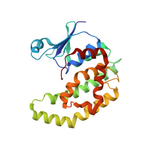 | |
UniProt | |||||
Entity Groups | |||||
| Sequence Clusters | 30% Identity50% Identity70% Identity90% Identity95% Identity100% Identity | ||||
| UniProt Group | Q5CCJ4 | ||||
Sequence AnnotationsExpand | |||||
Reference Sequence | |||||
| Ligands 2 Unique | |||||
|---|---|---|---|---|---|
| ID | Chains | Name / Formula / InChI Key | 2D Diagram | 3D Interactions | |
| GTS Download:Ideal Coordinates CCD File | C [auth A] | GLUTATHIONE SULFONIC ACID C10 H17 N3 O9 S QGWRMTHFAZVWAM-WDSKDSINSA-N |  | ||
| 1PE Download:Ideal Coordinates CCD File | B [auth A] | PENTAETHYLENE GLYCOL C10 H22 O6 JLFNLZLINWHATN-UHFFFAOYSA-N |  | ||
| Length ( Å ) | Angle ( ˚ ) |
|---|---|
| a = 57.41 | α = 90 |
| b = 57.41 | β = 90 |
| c = 209.665 | γ = 120 |
| Software Name | Purpose |
|---|---|
| HKL-2000 | data collection |
| PHASES | phasing |
| PHENIX | refinement |
| HKL-2000 | data reduction |
| SCALA | data scaling |


