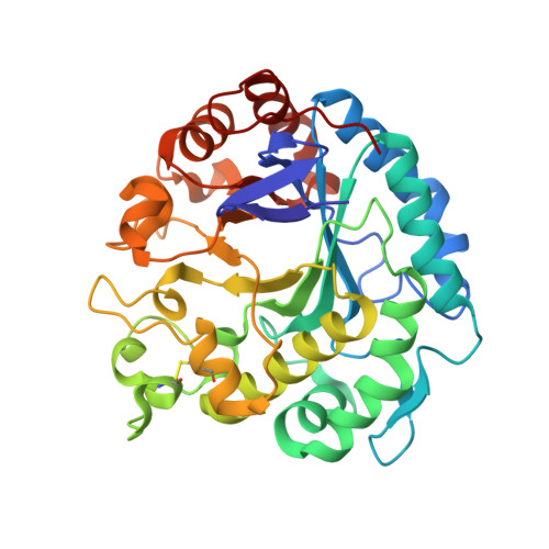
3VUP

Beta-1,4-mannanase from the common sea hare Aplysia kurodai


Experimental Data Snapshot

  • Method: X-RAY DIFFRACTION
  • Resolution: 1.05 Å
  • R-Value Free: 0.157 
  • R-Value Work: 0.137 
  • R-Value Observed: 0.138 

wwPDB Validation   3D Report Full Report


This is version 1.1 of the entry. See complete history


Literature

Structure of beta-1,4-mannanase from the common sea hare Aplysia kurodai at 1.05 A resolution.

Mizutani, K.Tsuchiya, S.Toyoda, M.Nanbu, Y.Tominaga, K.Yuasa, K.Takahashi, N.Tsuji, A.Mikami, B.

(2012) Acta Crystallogr Sect F Struct Biol Cryst Commun 68: 1164-1168

  • DOI: https://doi.org/10.1107/S1744309112037074
  • Primary Citation of Related Structures:  
    3VUP

  • PubMed Abstract: 

    β-1,4-Mannanase (EC 3.2.1.78) catalyzes the hydrolysis of β-1,4-glycosidic bonds within mannan, a major constituent group of the hemicelluloses. Bivalves and gastropods possess β-1,4-mannanase and may degrade mannan in seaweed and/or phytoplankton to obtain carbon and energy using the secreted enzymes in their digestive systems. In the present study, the crystal structure of AkMan, a gastropod β-1,4-mannanase prepared from the common sea hare Aplysia kurodai, was determined at 1.05 Å resolution. This is the first report of the three-dimensional structure of a gastropod β-1,4-mannanase. The structure was compared with bivalve β-1,4-mannanase and the roles of residues in the catalytic cleft were investigated. No obvious binding residue was found in subsite +1 and the substrate-binding site was exposed to the molecular surface, which may account for the enzymatic properties of mannanases that can digest complex substrates such as glucomannan and branched mannan.


  • Organizational Affiliation

    Laboratory of Applied Structural Biology, Division of Applied Life Sciences, Graduate School of Agriculture, Kyoto University, Uji, Kyoto 611-0011, Japan.


Macromolecules
Find similar proteins by:  (by identity cutoff)  |  3D Structure
Entity ID: 1
MoleculeChains Sequence LengthOrganismDetailsImage
Beta-1,4-mannanase
A, B
351Aplysia kurodaiMutation(s): 0 
UniProt
Find proteins for E5RSM0 (Aplysia kurodai)
Explore E5RSM0 
Go to UniProtKB:  E5RSM0
Entity Groups  
Sequence Clusters30% Identity50% Identity70% Identity90% Identity95% Identity100% Identity
UniProt GroupE5RSM0
Sequence Annotations
Expand
  • Reference Sequence
Small Molecules
Ligands 2 Unique
IDChains Name / Formula / InChI Key2D Diagram3D Interactions
TRS
Query on TRS

Download Ideal Coordinates CCD File 
C [auth A],
K [auth B]
2-AMINO-2-HYDROXYMETHYL-PROPANE-1,3-DIOL
C4 H12 N O3
LENZDBCJOHFCAS-UHFFFAOYSA-O
SO4
Query on SO4

Download Ideal Coordinates CCD File 
D [auth A]
E [auth A]
F [auth A]
G [auth A]
H [auth A]
D [auth A],
E [auth A],
F [auth A],
G [auth A],
H [auth A],
I [auth A],
J [auth A],
L [auth B],
M [auth B],
N [auth B],
O [auth B],
P [auth B],
Q [auth B],
R [auth B]
SULFATE ION
O4 S
QAOWNCQODCNURD-UHFFFAOYSA-L
Experimental Data & Validation

Experimental Data

  • Method: X-RAY DIFFRACTION
  • Resolution: 1.05 Å
  • R-Value Free: 0.157 
  • R-Value Work: 0.137 
  • R-Value Observed: 0.138 
  • Space Group: P 21 21 2
Unit Cell:
Length ( Å )Angle ( ˚ )
a = 110.092α = 90
b = 114.098β = 90
c = 51.138γ = 90
Software Package:
Software NamePurpose
HKL-2000data collection
MOLREPphasing
PHENIXrefinement
HKL-2000data reduction
HKL-2000data scaling

Structure Validation

View Full Validation Report


Entry History 

Deposition Data

Revision History  (Full details and data files)

  • Version 1.0: 2012-10-17
    Type: Initial release
  • Version 1.1: 2024-10-30
    Changes: Data collection, Database references, Derived calculations, Structure summary