3VPY | pdb_00003vpy

Crystal structure of Arabidopsis DDL FHA domain


Experimental Data Snapshot

  • Method: X-RAY DIFFRACTION
  • Resolution: 1.70 Å
  • R-Value Free: 
    0.231 (Depositor), 0.228 (DCC) 
  • R-Value Work: 
    0.188 (Depositor), 0.185 (DCC) 
  • R-Value Observed: 
    0.192 (Depositor) 

wwPDB Validation 3D Report Full Report

Validation slider image for 3VPY

This is version 1.1 of the entry. See complete history

Literature

Crystal Structure of Arabidopsis thaliana Dawdle Forkhead-Associated Domain reveals a conserved phospho-threonine recognition cleft for Dicer-like1 binding.

Machida, S.Yuan, A.Y.

(2013) Mol Plant 

  • DOI: https://doi.org/10.1093/mp/sst007
  • Primary Citation Related Structures: 
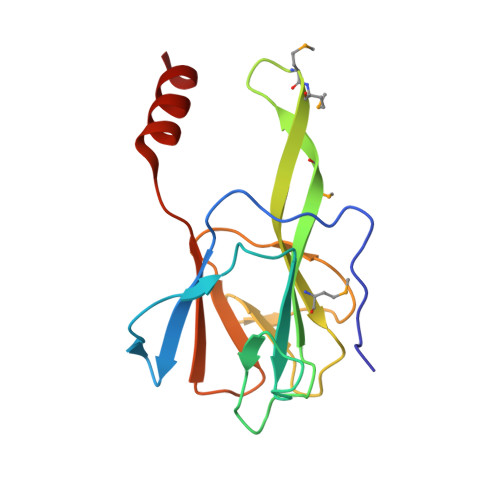
    3VPY

  • PubMed Abstract: 

    Dawdle (DDL) is a microRNA processing protein essential for the development of Arabidopsis. DDL contains a putative nuclear localization signal at its amino-terminus and forkhead-associated (FHA) domain at the carboxyl-terminus. Here, we report the crystal structure of the FHA domain of Arabidopsis Dawdle, determined by multiple-wavelength anomalous dispersion method at 1.7-Å resolution. DDL FHA structure displays a seven-stranded β-sandwich architecture that contains a unique structural motif comprising two long anti-parallel strands. Strikingly, crystal packing of the DDL FHA domain reveals that a glutamate residue from the symmetry-related DDL FHA domain, a structural mimic of the phospho-threonine, is specifically recognized by the structurally conserved phospho-threonine binding cleft. Consistently with the structural observations, co-immuno-precipitation experiments performed in Nicotiana benthamiana show that the DDL FHA domain co-immuno-precipitates with DCL1 fragments containing the predicted pThr+3(Ile/Val/Leu/Asp) motif. Taken together, we count the recognition of the target residue by the canonical binding cleft of the DDL FHA domain as the key molecular event to instate FHA domain-mediated protein-protein interaction in plant miRNA processing.


  • Organizational Affiliation
    • Department of Biological Sciences and Centre for Bioimaging Sciences, National University of Singapore, 14 Science Drive 4, Singapore 117543, Singapore.

Macromolecule Content 

  • Total Structure Weight: 17.53 kDa 
  • Atom Count: 1,302 
  • Modeled Residue Count: 145 
  • Deposited Residue Count: 145 
  • Unique protein chains: 1

Macromolecules

Find similar proteins by:|  3D Structure
Entity ID: 1
MoleculeChains  Sequence LengthOrganismDetailsImage
FHA domain-containing protein DDL145Arabidopsis thalianaMutation(s): 0 
Gene Names: DDLAt3g20550K10D20.9
UniProt
Find proteins for Q8W4D8 (Arabidopsis thaliana)
Explore Q8W4D8 
Go to UniProtKB:  Q8W4D8
Entity Groups
Sequence Clusters30% Identity50% Identity70% Identity90% Identity95% Identity100% Identity
UniProt GroupQ8W4D8
Sequence Annotations
Expand
Reference Sequence

Small Molecules

Modified Residues  1 Unique
IDChains TypeFormula2D DiagramParent
MSE
Query on MSE
A
L-PEPTIDE LINKINGC5 H11 N O2 SeMET

Experimental Data & Validation

Experimental Data

  • Method: X-RAY DIFFRACTION
  • Resolution: 1.70 Å
  • R-Value Free:  0.231 (Depositor), 0.228 (DCC) 
  • R-Value Work:  0.188 (Depositor), 0.185 (DCC) 
  • R-Value Observed: 0.192 (Depositor) 
Space Group: P 21 21 21
Unit Cell:
Length ( Å )Angle ( ˚ )
a = 48.564α = 90
b = 48.709β = 90
c = 54.688γ = 90
Software Package:
Software NamePurpose
HKL-2000data collection
SHARPphasing
REFMACrefinement
HKL-2000data reduction
HKL-2000data scaling

Structure Validation

View Full Validation Report


Entry History 

Deposition Data

Revision History  (Full details and data files)

  • Version 1.0: 2013-02-06
    Type: Initial release
  • Version 1.1: 2024-10-16
    Changes: Data collection, Database references, Derived calculations, Structure summary