3VOW | pdb_00003vow

Crystal Structure of the Human APOBEC3C having HIV-1 Vif-binding Interface


Experimental Data Snapshot

  • Method: X-RAY DIFFRACTION
  • Resolution: 2.15 Å
  • R-Value Free: 
    0.263 (Depositor), 0.269 (DCC) 
  • R-Value Work: 
    0.214 (Depositor) 
  • R-Value Observed: 
    0.216 (Depositor) 

Starting Model: experimental
View more details

wwPDB Validation 3D Report Full Report

Validation slider image for 3VOW

This is version 1.4 of the entry. See complete history

Literature

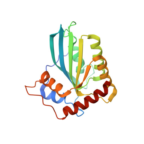
The APOBEC3C crystal structure and the interface for HIV-1 Vif binding.

Kitamura, S.Ode, H.Nakashima, M.Imahashi, M.Naganawa, Y.Kurosawa, T.Yokomaku, Y.Yamane, T.Watanabe, N.Suzuki, A.Sugiura, W.Iwatani, Y.

(2012) Nat Struct Mol Biol 19: 1005-1010

  • DOI: https://doi.org/10.1038/nsmb.2378
  • Primary Citation Related Structures: 
    3VOW

  • PubMed Abstract: 

    The human apolipoprotein B mRNA-editing enzyme catalytic polypeptide-like 3 (APOBEC3, referred to as A3) proteins are cellular cytidine deaminases that potently restrict retrovirus replication. However, HIV-1 viral infectivity factor (Vif) counteracts the antiviral activity of most A3 proteins by targeting them for proteasomal degradation. To date, the structure of an A3 protein containing a Vif-binding interface has not been solved. Here, we report a high-resolution crystal structure of APOBEC3C and identify the HIV-1 Vif-interaction interface. Extensive structure-guided mutagenesis revealed the role of a shallow cavity composed of hydrophobic or negatively charged residues between the α2 and α3 helices. This region is distant from the DPD motif (residues 128-130) of APOBEC3G that participates in HIV-1 Vif interaction. These findings provide insight into Vif-A3 interactions and could lead to the development of new pharmacologic anti-HIV-1 compounds.


  • Organizational Affiliation
    • Clinical Research Center, Department of Infectious Diseases and Immunology, National Hospital Organization Nagoya Medical Center, Nagoya, Japan.

Macromolecule Content 

  • Total Structure Weight: 45.87 kDa 
  • Atom Count: 3,281 
  • Modeled Residue Count: 372 
  • Deposited Residue Count: 380 
  • Unique protein chains: 1

Macromolecules

Find similar proteins by:|  3D Structure
Entity ID: 1
MoleculeChains  Sequence LengthOrganismDetailsImage
Probable DNA dC->dU-editing enzyme APOBEC-3C
A, B
190Homo sapiensMutation(s): 0 
Gene Names: APOBEC3CAPOBEC1LPBI
EC: 3.5.4 (PDB Primary Data), 3.5.4.38 (UniProt)
UniProt & NIH Common Fund Data Resources
Find proteins for Q9NRW3 (Homo sapiens)
Explore Q9NRW3 
Go to UniProtKB:  Q9NRW3
PHAROS:  Q9NRW3
GTEx:  ENSG00000244509 
Entity Groups
Sequence Clusters30% Identity50% Identity70% Identity90% Identity95% Identity100% Identity
UniProt GroupQ9NRW3
Sequence Annotations
Expand
Reference Sequence

Experimental Data & Validation

Experimental Data

  • Method: X-RAY DIFFRACTION
  • Resolution: 2.15 Å
  • R-Value Free:  0.263 (Depositor), 0.269 (DCC) 
  • R-Value Work:  0.214 (Depositor) 
  • R-Value Observed: 0.216 (Depositor) 
Space Group: P 61
Unit Cell:
Length ( Å )Angle ( ˚ )
a = 105.042α = 90
b = 105.042β = 90
c = 70.046γ = 120
Software Package:
Software NamePurpose
DENZOdata reduction
SCALEPACKdata scaling
MOLREPphasing
REFMACrefinement
PDB_EXTRACTdata extraction
ADSCdata collection
HKL-2000data reduction
HKL-2000data scaling

Structure Validation

View Full Validation Report


Entry History 

Deposition Data

Revision History  (Full details and data files)

  • Version 1.0: 2012-10-03
    Type: Initial release
  • Version 1.1: 2012-10-17
    Changes: Database references
  • Version 1.2: 2012-10-24
    Changes: Structure summary
  • Version 1.3: 2017-11-22
    Changes: Refinement description
  • Version 1.4: 2023-11-08
    Changes: Data collection, Database references, Derived calculations, Refinement description