3VOL | pdb_00003vol

X-ray Crystal Structure of PAS-HAMP Aer2 in the CN-bound Form


Experimental Data Snapshot

  • Method: X-RAY DIFFRACTION
  • Resolution: 2.40 Å
  • R-Value Free: 
    0.268 (Depositor), 0.263 (DCC) 
  • R-Value Work: 
    0.223 (Depositor), 0.220 (DCC) 
  • R-Value Observed: 
    0.225 (Depositor) 

wwPDB Validation 3D Report Full Report

Validation slider image for 3VOL

Ligand Structure Quality Assessment 


This is version 1.3 of the entry. See complete history

Literature

Structural basis for oxygen sensing and signal transduction of the heme-based sensor protein Aer2 from Pseudomonas aeruginosa

Sawai, H.Sugimoto, H.Shiro, Y.Ishikawa, H.Mizutani, Y.Aono, S.

(2012) Chem Commun (Camb) 48: 6523-6525

  • DOI: https://doi.org/10.1039/c2cc32549g
  • Primary Citation Related Structures: 
    3VOL

  • PubMed Abstract: 

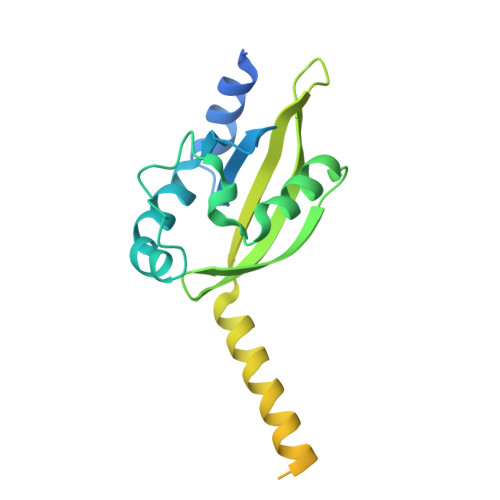
    The crystal structure of a truncated Aer2, a signal transducer protein from Pseudomonas aeruginosa, consisting of the heme-containing PAS and di-HAMP domains revealed that a distal tryptophan residue (Trp283) plays an important role in stabilizing the heme-bound O(2) and intra-molecular signal transduction upon O(2) binding.


  • Organizational Affiliation
    • Okazaki Institute for Integrative Bioscience, National Institutes of Natural Sciences, 5-1 Higashiyama, Myodaiji, Okazaki 444-8787, Japan.

Macromolecule Content 

  • Total Structure Weight: 26.44 kDa 
  • Atom Count: 1,123 
  • Modeled Residue Count: 138 
  • Deposited Residue Count: 233 
  • Unique protein chains: 1

Macromolecules

Find similar proteins by:|  3D Structure
Entity ID: 1
MoleculeChains  Sequence LengthOrganismDetailsImage
Aerotaxis transducer Aer2233Pseudomonas aeruginosa PAO1Mutation(s): 0 
Gene Names: aer2PA0176
UniProt
Find proteins for Q9I6V6 (Pseudomonas aeruginosa (strain ATCC 15692 / DSM 22644 / CIP 104116 / JCM 14847 / LMG 12228 / 1C / PRS 101 / PAO1))
Explore Q9I6V6 
Go to UniProtKB:  Q9I6V6
Entity Groups
Sequence Clusters30% Identity50% Identity70% Identity90% Identity95% Identity100% Identity
UniProt GroupQ9I6V6
Sequence Annotations
Expand
Reference Sequence

Small Molecules

Experimental Data & Validation

Experimental Data

  • Method: X-RAY DIFFRACTION
  • Resolution: 2.40 Å
  • R-Value Free:  0.268 (Depositor), 0.263 (DCC) 
  • R-Value Work:  0.223 (Depositor), 0.220 (DCC) 
  • R-Value Observed: 0.225 (Depositor) 
Space Group: P 65 2 2
Unit Cell:
Length ( Å )Angle ( ˚ )
a = 83.02α = 90
b = 83.02β = 90
c = 107.76γ = 120
Software Package:
Software NamePurpose
DENZOdata reduction
SCALEPACKdata scaling
MOLREPphasing
DMphasing
PHENIXrefinement
PDB_EXTRACTdata extraction
BSSdata collection
HKL-2000data reduction

Structure Validation

View Full Validation Report


Ligand Structure Quality Assessment 


Entry History 

Deposition Data

Revision History  (Full details and data files)

  • Version 1.0: 2012-05-23
    Type: Initial release
  • Version 1.1: 2013-07-31
    Changes: Database references
  • Version 1.2: 2017-11-22
    Changes: Refinement description
  • Version 1.3: 2024-03-20
    Changes: Data collection, Database references, Derived calculations