3VLZ | pdb_00003vlz

Assimilatory nitrite reductase (Nii3) - N226K mutant - SO3 full complex from tobacco leaf


Experimental Data Snapshot

  • Method: X-RAY DIFFRACTION
  • Resolution: 2.07 Å
  • R-Value Free: 
    0.282 (Depositor), 0.280 (DCC) 
  • R-Value Work: 
    0.223 (Depositor), 0.220 (DCC) 
  • R-Value Observed: 
    0.226 (Depositor) 

Starting Model: experimental
View more details

wwPDB Validation   3D Report Full Report


Ligand Structure Quality Assessment 

Created with Raphaël 2.3.0Worse 01 BetterLigand structure goodness of fit to experimental dataBest fitted SRMClick on this verticalbar to view details

This is version 2.0 of the entry. See complete history


Literature

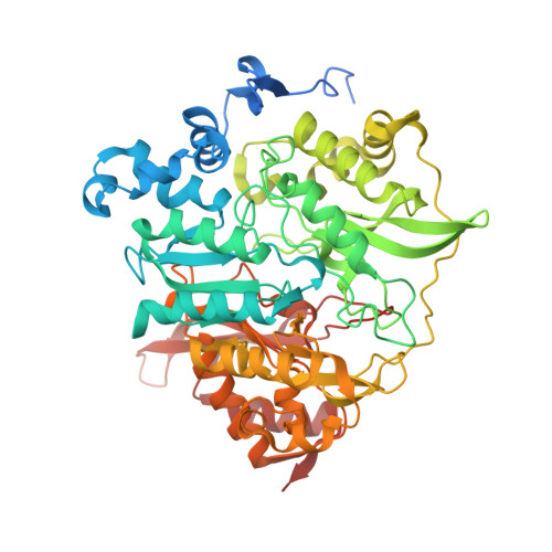
X-ray crystal structure of a mutant assimilatory nitrite reductase that shows sulfite reductase-like activity

Nakano, S.Takahashi, M.Sakamoto, A.Morikawa, H.Katayanagi, K.

(2012) Chem Biodivers 9: 1989-1999

  • DOI: https://doi.org/10.1002/cbdv.201100442
  • Primary Citation of Related Structures:  
    3VLX, 3VLY, 3VLZ, 3VM0, 3VM1

  • PubMed Abstract: 

    Assimilatory nitrite reductase (aNiR) reduces nitrite ions (NO(2)(-)) to ammonium ions (NH(4)(+)), whereas assimilatory sulfite reductase reduces sulfite (SO(3)(2-)) to hydrogen sulfide (HS(-)). Although aNiR can also reduce SO(3)(2-), its activity is much lower than when NO(2)(-) is reduced as the substrate. To increase the SO(3)(2-)-reduction activity of aNiR, we performed a N226K mutation of Nii3, a representative aNiR. The resulting Nii3-N226K variant could bind non-native targets, SO(3)(2-), and HCO(3)(-), in addition to its native target, i.e., NO(2)(-). We have determined the high-resolution structure of Nii3-N226K in its apo-state and in complex with SO(3)(2-), NO(2)(-), and HCO(3)(-). This analysis revealed conformational changes of Lys226 and the adjacent Lys224 upon binding of SO(3)(2-), but not NO(2)(-)In contrast, HCO(3)(-) binding induced a conformational change at Arg179. After replacing Asn226 with a positively charged Lys, aNiR showed affinity for several anions. A comparison of all ligand-bound structures for Nii3-N226K revealed that structural changes in the active site depend on the size of the substrate.


  • Organizational Affiliation

    Department of Mathematical and Life Sciences, Graduate School of Science, Hiroshima University, 1-3-1, Kagamiyama, Higashi-Hiroshima 739-8526, Japan.


Macromolecules
Find similar proteins by:  (by identity cutoff)  |  3D Structure
Entity ID: 1
MoleculeChains Sequence LengthOrganismDetailsImage
Nitrite reductase584Nicotiana tabacumMutation(s): 1 
Gene Names: nii3
EC: 1.7.7.1
UniProt
Find proteins for Q76KB0 (Nicotiana tabacum)
Explore Q76KB0 
Go to UniProtKB:  Q76KB0
Entity Groups  
Sequence Clusters30% Identity50% Identity70% Identity90% Identity95% Identity100% Identity
UniProt GroupQ76KB0
Sequence Annotations
Expand
  • Reference Sequence
Small Molecules
Ligands 5 Unique
IDChains Name / Formula / InChI Key2D Diagram3D Interactions
SRM
Query on SRM

Download Ideal Coordinates CCD File 
B [auth A]SIROHEME
C42 H44 Fe N4 O16
DLKSSIHHLYNIKN-MWBYXLBFSA-L
SF4
Query on SF4

Download Ideal Coordinates CCD File 
C [auth A]IRON/SULFUR CLUSTER
Fe4 S4
LJBDFODJNLIPKO-UHFFFAOYSA-N
SO3
Query on SO3

Download Ideal Coordinates CCD File 
G [auth A]SULFITE ION
O3 S
LSNNMFCWUKXFEE-UHFFFAOYSA-L
K
Query on K

Download Ideal Coordinates CCD File 
D [auth A]POTASSIUM ION
K
NPYPAHLBTDXSSS-UHFFFAOYSA-N
CL
Query on CL

Download Ideal Coordinates CCD File 
E [auth A],
F [auth A]
CHLORIDE ION
Cl
VEXZGXHMUGYJMC-UHFFFAOYSA-M
Experimental Data & Validation

Experimental Data

  • Method: X-RAY DIFFRACTION
  • Resolution: 2.07 Å
  • R-Value Free:  0.282 (Depositor), 0.280 (DCC) 
  • R-Value Work:  0.223 (Depositor), 0.220 (DCC) 
  • R-Value Observed: 0.226 (Depositor) 
Space Group: P 4 21 2
Unit Cell:
Length ( Å )Angle ( ˚ )
a = 132.889α = 90
b = 132.889β = 90
c = 77.646γ = 90
Software Package:
Software NamePurpose
SCALEPACKdata scaling
MOLREPphasing
REFMACrefinement
PDB_EXTRACTdata extraction
ADSCdata collection
HKL-2000data reduction

Structure Validation

View Full Validation Report


Ligand Structure Quality Assessment 

Created with Raphaël 2.3.0Worse 01 BetterLigand structure goodness of fit to experimental dataBest fitted SRMClick on this verticalbar to view details

Entry History 

Deposition Data

Revision History  (Full details and data files)

  • Version 1.0: 2012-09-26
    Type: Initial release
  • Version 1.1: 2023-11-08
    Changes: Data collection, Database references, Derived calculations, Refinement description
  • Version 2.0: 2023-12-27
    Changes: Data collection, Non-polymer description, Structure summary