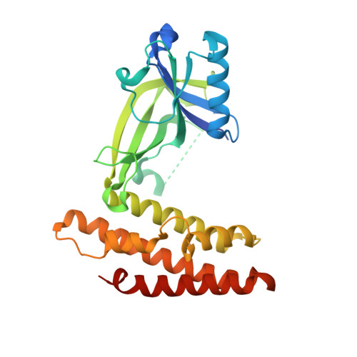
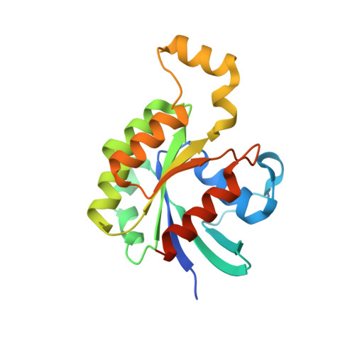
3VHL

Crystal structure of the DHR-2 domain of DOCK8 in complex with Cdc42 (T17N mutant)


Experimental Data Snapshot

  • Method: X-RAY DIFFRACTION
  • Resolution: 2.08 Å
  • R-Value Free: 0.232 
  • R-Value Work: 0.178 
  • R-Value Observed: 0.182 

Starting Model: experimental
View more details

wwPDB Validation   3D Report Full Report


This is version 1.1 of the entry. See complete history


Literature

DOCK8 is a Cdc42 activator critical for interstitial dendritic cell migration during immune responses.

Harada, Y.Tanaka, Y.Terasawa, M.Pieczyk, M.Habiro, K.Katakai, T.Hanawa-Suetsugu, K.Kukimoto-Niino, M.Nishizaki, T.Shirouzu, M.Duan, X.Uruno, T.Nishikimi, A.Sanematsu, F.Yokoyama, S.Stein, J.V.Kinashi, T.Fukui, Y.

(2012) Blood 119: 4451-4461

  • DOI: https://doi.org/10.1182/blood-2012-01-407098
  • Primary Citation of Related Structures:  
    3VHL

  • PubMed Abstract: 

    To migrate efficiently through the interstitium, dendritic cells (DCs) constantly adapt their shape to the given structure of the extracellular matrix and follow the path of least resistance. It is known that this amoeboid migration of DCs requires Cdc42, yet the upstream regulators critical for localization and activation of Cdc42 remain to be determined. Mutations of DOCK8, a member of the atypical guanine nucleotide exchange factor family, causes combined immunodeficiency in humans. In the present study, we show that DOCK8 is a Cdc42-specific guanine nucleotide exchange factor that is critical for interstitial DC migration. By generating the knockout mice, we found that in the absence of DOCK8, DCs failed to accumulate in the lymph node parenchyma for T-cell priming. Although DOCK8-deficient DCs migrated normally on 2-dimensional surfaces, DOCK8 was required for DCs to crawl within 3-dimensional fibrillar networks and to transmigrate through the subcapsular sinus floor. This function of DOCK8 depended on the DHR-2 domain mediating Cdc42 activation. DOCK8 deficiency did not affect global Cdc42 activity. However, Cdc42 activation at the leading edge membrane was impaired in DOCK8-deficient DCs, resulting in a severe defect in amoeboid polarization and migration. Therefore, DOCK8 regulates interstitial DC migration by controlling Cdc42 activity spatially.


  • Organizational Affiliation

    Division of Immunogenetics, Department of Immunobiology and Neuroscience, Medical Institute of Bioregulation, Kyushu University, Fukuoka, Japan.


Macromolecules
Find similar proteins by:  (by identity cutoff)  |  3D Structure
Entity ID: 1
MoleculeChains Sequence LengthOrganismDetailsImage
Dedicator of cytokinesis protein 8288Mus musculusMutation(s): 0 
Gene Names: Dock8
UniProt
Find proteins for Q8C147 (Mus musculus)
Explore Q8C147 
Go to UniProtKB:  Q8C147
Entity Groups  
Sequence Clusters30% Identity50% Identity70% Identity90% Identity95% Identity100% Identity
UniProt GroupQ8C147
Sequence Annotations
Expand
  • Reference Sequence
Find similar proteins by:  (by identity cutoff)  |  3D Structure
Entity ID: 2
MoleculeChains Sequence LengthOrganismDetailsImage
Cell division control protein 42 homolog195Homo sapiensMutation(s): 2 
Gene Names: CDC42
EC: 3.6.5.2
UniProt & NIH Common Fund Data Resources
Find proteins for P60953 (Homo sapiens)
Explore P60953 
Go to UniProtKB:  P60953
PHAROS:  P60953
GTEx:  ENSG00000070831 
Entity Groups  
Sequence Clusters30% Identity50% Identity70% Identity90% Identity95% Identity100% Identity
UniProt GroupP60953
Sequence Annotations
Expand
  • Reference Sequence
Experimental Data & Validation

Experimental Data

  • Method: X-RAY DIFFRACTION
  • Resolution: 2.08 Å
  • R-Value Free: 0.232 
  • R-Value Work: 0.178 
  • R-Value Observed: 0.182 
  • Space Group: C 1 2 1
Unit Cell:
Length ( Å )Angle ( ˚ )
a = 104.185α = 90
b = 73.279β = 104.82
c = 65.031γ = 90
Software Package:
Software NamePurpose
PHENIXmodel building
PHENIXrefinement
HKL-2000data reduction
HKL-2000data scaling
PHENIXphasing

Structure Validation

View Full Validation Report


Entry History 

Deposition Data

Revision History  (Full details and data files)

  • Version 1.0: 2012-06-20
    Type: Initial release
  • Version 1.1: 2023-11-08
    Changes: Data collection, Database references, Derived calculations, Refinement description