


Crystal structure of orotidine 5'-monophosphate decarboxylase from Metallosphaera sedula complexed with inhibitor BMP
Fedorov, A.A., Fedorov, E.V., Desai, B., Gerlt, J.A., Almo, S.C.To be published.
Experimental Data Snapshot
Entity ID: 1 | |||||
|---|---|---|---|---|---|
| Molecule | Chains | Sequence Length | Organism | Details | Image |
| Orotidine-5'-phosphate decarboxylase | 215 | Metallosphaera sedula DSM 5348 | Mutation(s): 0 Gene Names: Msed_1966 EC: 4.1.1.23 | 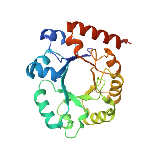 | |
UniProt | |||||
Entity Groups | |||||
| Sequence Clusters | 30% Identity50% Identity70% Identity90% Identity95% Identity100% Identity | ||||
| UniProt Group | A4YI54 | ||||
Sequence AnnotationsExpand | |||||
Reference Sequence | |||||
| Ligands 3 Unique | |||||
|---|---|---|---|---|---|
| ID | Chains | Name / Formula / InChI Key | 2D Diagram | 3D Interactions | |
| BMP Download:Ideal Coordinates CCD File | C [auth A], E [auth B] | 6-HYDROXYURIDINE-5'-PHOSPHATE C9 H13 N2 O10 P UDOBICLZEKUKCV-YXZULKJRSA-N |  | ||
| SO4 Download:Ideal Coordinates CCD File | D [auth A], I [auth B], J [auth B] | SULFATE ION O4 S QAOWNCQODCNURD-UHFFFAOYSA-L |  | ||
| ACY Download:Ideal Coordinates CCD File | F [auth B], G [auth B], H [auth B] | ACETIC ACID C2 H4 O2 QTBSBXVTEAMEQO-UHFFFAOYSA-N |  | ||
| Length ( Å ) | Angle ( ˚ ) |
|---|---|
| a = 41.396 | α = 90 |
| b = 91.57 | β = 98.79 |
| c = 50.954 | γ = 90 |
| Software Name | Purpose |
|---|---|
| CBASS | data collection |
| PHENIX | model building |
| PHENIX | refinement |
| HKL-2000 | data reduction |
| SCALEPACK | data scaling |
| PHENIX | phasing |


