3VDL | pdb_00003vdl

Crystal structure of circumsporozoite protein aTSR domain, P43212 form


Experimental Data Snapshot

  • Method: X-RAY DIFFRACTION
  • Resolution: 2.04 Å
  • R-Value Free: 
    0.240 (Depositor), 0.242 (DCC) 
  • R-Value Work: 
    0.194 (Depositor), 0.195 (DCC) 
  • R-Value Observed: 
    0.196 (Depositor) 

Starting Model: experimental
View more details

wwPDB Validation 3D Report Full Report

Validation slider image for 3VDL

This is version 1.4 of the entry. See complete history

Literature

Unexpected fold in the circumsporozoite protein target of malaria vaccines.

Doud, M.B.Koksal, A.C.Mi, L.Z.Song, G.Lu, C.Springer, T.A.

(2012) Proc Natl Acad Sci U S A 109: 7817-7822

  • DOI: https://doi.org/10.1073/pnas.1205737109
  • Primary Citation Related Structures: 
    3VDJ, 3VDK, 3VDL

  • PubMed Abstract: 

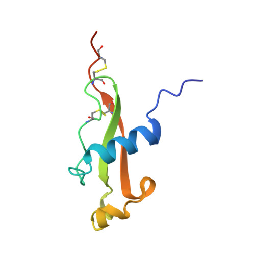
    Circumsporozoite (CS) protein is the major surface component of Plasmodium falciparum sporozoites and is essential for host cell invasion. A vaccine containing tandem repeats, region III, and thrombospondin type-I repeat (TSR) of CS is efficacious in phase III trials but gives only a 35% reduction in severe malaria in the first year postimmunization. We solved crystal structures showing that region III and TSR fold into a single unit, an "αTSR" domain. The αTSR domain possesses a hydrophobic pocket and core, missing in TSR domains. CS binds heparin, but αTSR does not. Interestingly, polymorphic T-cell epitopes map to specialized αTSR regions. The N and C termini are unexpectedly close, providing clues for sporozoite sheath organization. Elucidation of a unique structure of a domain within CS enables rational design of next-generation subunit vaccines and functional and medicinal chemical investigation of the conserved hydrophobic pocket.


  • Organizational Affiliation
    • Immune Disease Institute, Children's Hospital Boston and Department of Biological Chemistry and Molecular Pharmacology, Harvard Medical School, Boston, MA 02115, USA.

Macromolecule Content 

  • Total Structure Weight: 26.71 kDa 
  • Atom Count: 1,791 
  • Modeled Residue Count: 213 
  • Deposited Residue Count: 231 
  • Unique protein chains: 1

Macromolecules

Find similar proteins by:|  3D Structure
Entity ID: 1
MoleculeChains  Sequence LengthOrganismDetailsImage
Circumsporozoite (CS) protein
A, B, C
77Plasmodium falciparum 3D7Mutation(s): 0 
Gene Names: PFC0210c
UniProt
Find proteins for Q7K740 (Plasmodium falciparum (isolate 3D7))
Explore Q7K740 
Go to UniProtKB:  Q7K740
Entity Groups
Sequence Clusters30% Identity50% Identity70% Identity90% Identity95% Identity100% Identity
UniProt GroupQ7K740
Sequence Annotations
Expand
Reference Sequence

Experimental Data & Validation

Experimental Data

  • Method: X-RAY DIFFRACTION
  • Resolution: 2.04 Å
  • R-Value Free:  0.240 (Depositor), 0.242 (DCC) 
  • R-Value Work:  0.194 (Depositor), 0.195 (DCC) 
  • R-Value Observed: 0.196 (Depositor) 
Space Group: P 43 21 2
Unit Cell:
Length ( Å )Angle ( ˚ )
a = 63.372α = 90
b = 63.372β = 90
c = 118.164γ = 90
Software Package:
Software NamePurpose
ADSCdata collection
PHASERphasing
PHENIXrefinement
HKL-2000data reduction
HKL-2000data scaling

Structure Validation

View Full Validation Report


Entry History 

Deposition Data

Revision History  (Full details and data files)

  • Version 1.0: 2012-05-09
    Type: Initial release
  • Version 1.1: 2012-05-16
    Changes: Database references
  • Version 1.2: 2012-05-30
    Changes: Database references
  • Version 1.3: 2023-09-13
    Changes: Data collection, Database references, Refinement description
  • Version 1.4: 2024-10-16
    Changes: Structure summary