3VCY | pdb_00003vcy

Structure of MurA (UDP-N-acetylglucosamine enolpyruvyl transferase), from Vibrio fischeri in complex with substrate UDP-N-acetylglucosamine and the drug fosfomycin.


Experimental Data Snapshot

  • Method: X-RAY DIFFRACTION
  • Resolution: 1.93 Å
  • R-Value Free: 
    0.261 (Depositor), 0.264 (DCC) 
  • R-Value Work: 
    0.199 (Depositor), 0.202 (DCC) 
  • R-Value Observed: 
    0.202 (Depositor) 

Starting Model: experimental
View more details

wwPDB Validation 3D Report Full Report

Validation slider image for 3VCY

Ligand Structure Quality Assessment 


This is version 1.3 of the entry. See complete history

Literature

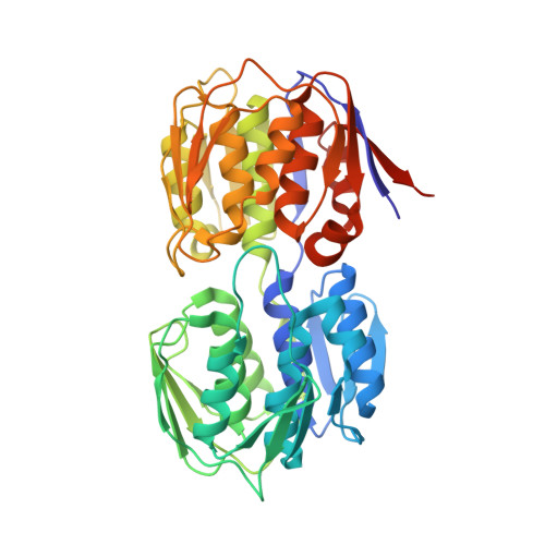
Structure of MurA (UDP-N-acetylglucosamine enolpyruvyl transferase) from Vibrio fischeri in complex with substrate UDP-N-acetylglucosamine and the drug fosfomycin.

Bensen, D.C.Rodriguez, S.Nix, J.Cunningham, M.L.Tari, L.W.

(2012) Acta Crystallogr Sect F Struct Biol Cryst Commun 68: 382-385

  • DOI: https://doi.org/10.1107/S1744309112006720
  • Primary Citation Related Structures: 
    3VCY

  • PubMed Abstract: 

    The development of new antibiotics is necessitated by the rapid development of resistance to current therapies. UDP-N-acetylglucosamine enolpyruvyl transferase (MurA), which catalyzes the first committed step of bacterial peptidoglycan biosynthesis, is a prime candidate for therapeutic intervention. MurA is the target of the antibiotic fosfomycin, a natural product produced by Streptomyces. Despite possessing a high degree of sequence conservation with MurA enzymes from fosfomycin-susceptible organisms, recent microbiological studies suggest that MurA from Vibrio fischeri (VfiMurA) may confer fosfomycin resistance via a mechanism that is not yet understood. The crystal structure of VfiMurA in a ternary complex with the substrate UDP-N-acetylglucosamine (UNAG) and fosfomycin has been solved to a resolution of 1.93 Å. Fosfomycin is known to inhibit MurA by covalently binding to a highly conserved cysteine in the active site of the enzyme. A comparison of the title structure with the structure of fosfomycin-susceptible Haemophilus influenzae MurA (PDB entry 2rl2) revealed strikingly similar conformations of the mobile substrate-binding loop and clear electron density for a fosfomycin-cysteine adduct. Based on these results, there are no distinguishing sequence/structural features in VfiMurA that would translate to a diminished sensitivity to fosfomycin. However, VfiMurA is a robust crystallizer and shares high sequence identity with many clinically relevant bacterial pathogens. Thus, it would serve as an ideal system for use in the structure-guided optimization of new antibacterial agents.


  • Organizational Affiliation
    • Structural Biology, Trius Therapeutics, 6310 Nancy Ridge Drive, Suite 101, San Diego, CA 92008, USA.

Macromolecule Content 

  • Total Structure Weight: 186.94 kDa 
  • Atom Count: 13,938 
  • Modeled Residue Count: 1,676 
  • Deposited Residue Count: 1,720 
  • Unique protein chains: 1

Macromolecules

Find similar proteins by:|  3D Structure
Entity ID: 1
MoleculeChains  Sequence LengthOrganismDetailsImage
UDP-N-acetylglucosamine 1-carboxyvinyltransferase
A, B, C, D
430Aliivibrio fischeri MJ11Mutation(s): 0 
Gene Names: murAVFMJ11_0391
EC: 2.5.1.7
UniProt
Find proteins for B5F9P4 (Aliivibrio fischeri (strain MJ11))
Explore B5F9P4 
Go to UniProtKB:  B5F9P4
Entity Groups
Sequence Clusters30% Identity50% Identity70% Identity90% Identity95% Identity100% Identity
UniProt GroupB5F9P4
Sequence Annotations
Expand
Reference Sequence

Small Molecules

Ligands 4 Unique
IDChains Name / Formula / InChI Key2D Diagram3D Interactions
UD1

Query on UD1


Download:Ideal Coordinates CCD File
F [auth A],
I [auth B],
M [auth C],
Q [auth D]
URIDINE-DIPHOSPHATE-N-ACETYLGLUCOSAMINE
C17 H27 N3 O17 P2
LFTYTUAZOPRMMI-CFRASDGPSA-N
FFQ

Query on FFQ


Download:Ideal Coordinates CCD File
E [auth A],
J [auth B],
O [auth C],
T [auth D]
[(1R)-1-hydroxypropyl]phosphonic acid
C3 H9 O4 P
MVIJUJBSAAUHEM-GSVOUGTGSA-N
PO4

Query on PO4


Download:Ideal Coordinates CCD File
G [auth A],
K [auth B],
N [auth C],
R [auth D]
PHOSPHATE ION
O4 P
NBIIXXVUZAFLBC-UHFFFAOYSA-K
GOL

Query on GOL


Download:Ideal Coordinates CCD File
H [auth A],
L [auth B],
P [auth C],
S [auth D]
GLYCEROL
C3 H8 O3
PEDCQBHIVMGVHV-UHFFFAOYSA-N

Experimental Data & Validation

Experimental Data

  • Method: X-RAY DIFFRACTION
  • Resolution: 1.93 Å
  • R-Value Free:  0.261 (Depositor), 0.264 (DCC) 
  • R-Value Work:  0.199 (Depositor), 0.202 (DCC) 
  • R-Value Observed: 0.202 (Depositor) 
Space Group: P 1 21 1
Unit Cell:
Length ( Å )Angle ( ˚ )
a = 86.6α = 90
b = 118.51β = 116.48
c = 92.51γ = 90
Software Package:
Software NamePurpose
CrystalCleardata collection
PHASERphasing
REFMACrefinement
CrystalCleardata reduction
SCALAdata scaling

Structure Validation

View Full Validation Report


Ligand Structure Quality Assessment 


Entry History 

Deposition Data

Revision History  (Full details and data files)

  • Version 1.0: 2012-04-11
    Type: Initial release
  • Version 1.1: 2012-04-25
    Changes: Database references
  • Version 1.2: 2023-09-13
    Changes: Data collection, Database references, Derived calculations, Refinement description
  • Version 1.3: 2024-10-30
    Changes: Structure summary