


Crystal structure of Staphylococcal GAPDH1 in a hexagonal space group
Roychowdhury, A., Mukherjee, S., Dutta, D., Das, A.K.To be published.
Experimental Data Snapshot
Starting Model: experimental
View more details
Entity ID: 1 | |||||
|---|---|---|---|---|---|
| Molecule | Chains | Sequence Length | Organism | Details | Image |
| Glyceraldehyde-3-phosphate dehydrogenase 1 | 344 | Staphylococcus aureus subsp. aureus MRSA252 | Mutation(s): 0 Gene Names: gap, gapA, gapA1, SAR0828 EC: 1.2.1.12 | 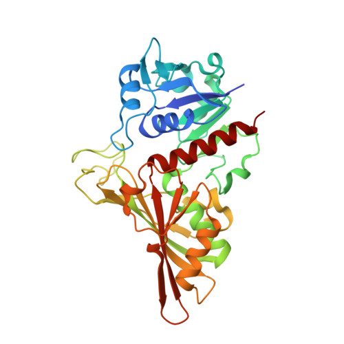 | |
UniProt | |||||
Entity Groups | |||||
| Sequence Clusters | 30% Identity50% Identity70% Identity90% Identity95% Identity100% Identity | ||||
| UniProt Group | Q6GIL8 | ||||
Sequence AnnotationsExpand | |||||
Reference Sequence | |||||
| Ligands 3 Unique | |||||
|---|---|---|---|---|---|
| ID | Chains | Name / Formula / InChI Key | 2D Diagram | 3D Interactions | |
| NAD Download:Ideal Coordinates CCD File | G [auth P] K [auth R] L [auth O] O [auth Q] P [auth B] | NICOTINAMIDE-ADENINE-DINUCLEOTIDE C21 H27 N7 O14 P2 BAWFJGJZGIEFAR-NNYOXOHSSA-N |  | ||
| DGY Download:Ideal Coordinates CCD File | N [auth O], T [auth A] | (2R)-2,3-DIHYDROXYPROPANOIC ACID C3 H6 O4 RBNPOMFGQQGHHO-UWTATZPHSA-N |  | ||
| PO4 Download:Ideal Coordinates CCD File | H [auth P] I [auth P] J [auth P] M [auth O] Q [auth B] | PHOSPHATE ION O4 P NBIIXXVUZAFLBC-UHFFFAOYSA-K |  | ||
| Length ( Å ) | Angle ( ˚ ) |
|---|---|
| a = 155.389 | α = 90 |
| b = 155.389 | β = 90 |
| c = 317.849 | γ = 120 |
| Software Name | Purpose |
|---|---|
| XSCALE | data scaling |
| PHASER | phasing |
| REFMAC | refinement |
| PDB_EXTRACT | data extraction |
| XDS | data reduction |
| XDS | data scaling |


