3VAU | pdb_00003vau

Myoglobin nitrite structure: nitriheme modified


Experimental Data Snapshot

  • Method: X-RAY DIFFRACTION
  • Resolution: 1.70 Å
  • R-Value Free: 
    0.237 (Depositor), 0.230 (DCC) 
  • R-Value Work: 
    0.173 (Depositor), 0.170 (DCC) 
  • R-Value Observed: 
    0.176 (Depositor) 

Starting Model: experimental
View more details

wwPDB Validation 3D Report Full Report

Validation slider image for 3VAU

Ligand Structure Quality Assessment 


This is version 1.1 of the entry. See complete history

Literature

Unveiling the three-dimensional structure of the green pigment of nitrite-cured meat

Yi, J.Richter-Addo, G.B.

(2012) Chem Commun (Camb) 48: 4172-4174

  • DOI: https://doi.org/10.1039/c2cc31065a
  • Primary Citation Related Structures: 
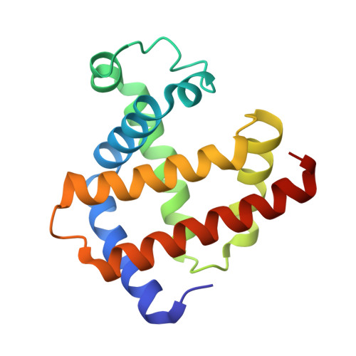
    3VAU

  • PubMed Abstract: 

    The 1.70 Å-resolution X-ray crystal structure of the green pigment occasionally present in nitrite-cured meat reveals a regiospecific nitration of the heme at the 2-vinyl position; the regiospecificity is favored on steric grounds, and results in movement of the 2-nitrovinyl moiety into the plane of the 24-atom heme macrocycle.


  • Organizational Affiliation
    • Department of Chemistry and Biochemistry, University of Oklahoma, Norman, Oklahoma, USA. yijun@ou.edu

Macromolecule Content 

  • Total Structure Weight: 17.79 kDa 
  • Atom Count: 1,336 
  • Modeled Residue Count: 151 
  • Deposited Residue Count: 153 
  • Unique protein chains: 1

Macromolecules

Find similar proteins by:|  3D Structure
Entity ID: 1
MoleculeChains  Sequence LengthOrganismDetailsImage
Myoglobin153Equus caballusMutation(s): 0 
EC: 1.11.1 (UniProt), 1.7 (UniProt)
UniProt
Find proteins for P68082 (Equus caballus)
Explore P68082 
Go to UniProtKB:  P68082
Entity Groups
Sequence Clusters30% Identity50% Identity70% Identity90% Identity95% Identity100% Identity
UniProt GroupP68082
Sequence Annotations
Expand
Reference Sequence

Small Molecules

Ligands 3 Unique
IDChains Name / Formula / InChI Key2D Diagram3D Interactions
NTE

Query on NTE


Download:Ideal Coordinates CCD File
B [auth A][3,3'-{7-ethenyl-3,8,13,17-tetramethyl-12-[(E)-2-nitroethenyl]porphyrin-2,18-diyl-kappa~4~N~21~,N~22~,N~23~,N~24~}dipro panoato(2-)]iron
C34 H31 Fe N5 O6
MBEUICWDKPMPCN-RXJYIKGQSA-L
SO4

Query on SO4


Download:Ideal Coordinates CCD File
D [auth A]SULFATE ION
O4 S
QAOWNCQODCNURD-UHFFFAOYSA-L
NO2

Query on NO2


Download:Ideal Coordinates CCD File
C [auth A]NITRITE ION
N O2
IOVCWXUNBOPUCH-UHFFFAOYSA-M

Experimental Data & Validation

Experimental Data

  • Method: X-RAY DIFFRACTION
  • Resolution: 1.70 Å
  • R-Value Free:  0.237 (Depositor), 0.230 (DCC) 
  • R-Value Work:  0.173 (Depositor), 0.170 (DCC) 
  • R-Value Observed: 0.176 (Depositor) 
Space Group: P 1 21 1
Unit Cell:
Length ( Å )Angle ( ˚ )
a = 35.14α = 90
b = 28.423β = 105.5
c = 62.867γ = 90
Software Package:
Software NamePurpose
CrystalCleardata collection
PHASERphasing
REFMACrefinement
d*TREKdata reduction
d*TREKdata scaling

Structure Validation

View Full Validation Report


Ligand Structure Quality Assessment 


Entry History 

Deposition Data

Revision History  (Full details and data files)

  • Version 1.0: 2012-04-11
    Type: Initial release
  • Version 1.1: 2023-11-08
    Changes: Data collection, Database references, Derived calculations, Refinement description, Structure summary