3V8E | pdb_00003v8e

Crystal structure of the yeast nicotinamidase Pnc1p bound to the inhibitor nicotinaldehyde


Experimental Data Snapshot

  • Method: X-RAY DIFFRACTION
  • Resolution: 2.71 Å
  • R-Value Free: 
    0.203 (Depositor), 0.202 (DCC) 
  • R-Value Work: 
    0.181 (Depositor), 0.180 (DCC) 
  • R-Value Observed: 
    0.182 (Depositor) 

wwPDB Validation 3D Report Full Report

Validation slider image for 3V8E

This is version 1.1 of the entry. See complete history

Literature

Structural and Kinetic Isotope Effect Studies of Nicotinamidase (Pnc1) from Saccharomyces cerevisiae.

Smith, B.C.Anderson, M.A.Hoadley, K.A.Keck, J.L.Cleland, W.W.Denu, J.M.

(2012) Biochemistry 51: 243-256

  • DOI: https://doi.org/10.1021/bi2015508
  • Primary Citation Related Structures: 
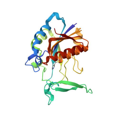
    3V8E

  • PubMed Abstract: 

    Nicotinamidases catalyze the hydrolysis of nicotinamide to nicotinic acid and ammonia. Nicotinamidases are absent in mammals but function in NAD(+) salvage in many bacteria, yeast, plants, protozoa, and metazoans. We have performed structural and kinetic investigations of the nicotinamidase from Saccharomyces cerevisiae (Pnc1). Steady-state product inhibitor analysis revealed an irreversible reaction in which ammonia is the first product released, followed by nicotinic acid. A series of nicotinamide analogues acting as inhibitors or substrates were examined, revealing that the nicotinamide carbonyl oxygen and ring nitrogen are critical for binding and reactivity. X-ray structural analysis revealed a covalent adduct between nicotinaldehyde and Cys167 of Pnc1 and coordination of the nicotinamide ring nitrogen to the active-site zinc ion. Using this structure as a guide, the function of several residues was probed via mutagenesis and primary (15)N and (13)C kinetic isotope effects (KIEs) on V/K for amide bond hydrolysis. The KIE values of almost all variants were increased, indicating that C-N bond cleavage is at least partially rate limiting; however, a decreased KIE for D51N was indicative of a stronger commitment to catalysis. In addition, KIE values using slower alternate substrates indicated that C-N bond cleavage is at least partially rate limiting with nicotinamide to highly rate limiting with thionicotinamide. A detailed mechanism involving nucleophilic attack of Cys167, followed by elimination of ammonia and then hydrolysis to liberate nicotinic acid, is discussed. These results will aid in the design of mechanism-based inhibitors to target pathogens that rely on nicotinamidase activity.


  • Organizational Affiliation
    • Department of Biomolecular Chemistry, University of Wisconsin, Madison, Wisconsin 53706, United States.

Macromolecule Content 

  • Total Structure Weight: 176.58 kDa 
  • Atom Count: 12,745 
  • Modeled Residue Count: 1,512 
  • Deposited Residue Count: 1,512 
  • Unique protein chains: 1

Macromolecules

Find similar proteins by:|  3D Structure
Entity ID: 1
MoleculeChains  Sequence LengthOrganismDetailsImage
Nicotinamidase
A, B, C, D, E
A, B, C, D, E, F, G
216Saccharomyces cerevisiae S288CMutation(s): 0 
Gene Names: PNC1YGL037C
EC: 3.5.1.19
UniProt
Find proteins for P53184 (Saccharomyces cerevisiae (strain ATCC 204508 / S288c))
Explore P53184 
Go to UniProtKB:  P53184
Entity Groups
Sequence Clusters30% Identity50% Identity70% Identity90% Identity95% Identity100% Identity
UniProt GroupP53184
Sequence Annotations
Expand
Reference Sequence

Small Molecules

Ligands 2 Unique
IDChains Name / Formula / InChI Key2D Diagram3D Interactions
ZN

Query on ZN


Download:Ideal Coordinates CCD File
H [auth A]
J [auth B]
L [auth C]
N [auth D]
P [auth E]
H [auth A],
J [auth B],
L [auth C],
N [auth D],
P [auth E],
R [auth F],
T [auth G]
ZINC ION
Zn
PTFCDOFLOPIGGS-UHFFFAOYSA-N
MG

Query on MG


Download:Ideal Coordinates CCD File
I [auth A]
K [auth B]
M [auth C]
O [auth D]
Q [auth E]
I [auth A],
K [auth B],
M [auth C],
O [auth D],
Q [auth E],
S [auth F],
U [auth G]
MAGNESIUM ION
Mg
JLVVSXFLKOJNIY-UHFFFAOYSA-N
Modified Residues  1 Unique
IDChains TypeFormula2D DiagramParent
JJJ
Query on JJJ
A, B, C, D, E
A, B, C, D, E, F, G
L-PEPTIDE LINKINGC9 H10 N2 O3 SCYS

Experimental Data & Validation

Experimental Data

  • Method: X-RAY DIFFRACTION
  • Resolution: 2.71 Å
  • R-Value Free:  0.203 (Depositor), 0.202 (DCC) 
  • R-Value Work:  0.181 (Depositor), 0.180 (DCC) 
  • R-Value Observed: 0.182 (Depositor) 
Space Group: H 3
Unit Cell:
Length ( Å )Angle ( ˚ )
a = 298.717α = 90
b = 298.717β = 90
c = 112.652γ = 120
Software Package:
Software NamePurpose
REFMACrefinement

Structure Validation

View Full Validation Report


Entry History 

Deposition Data

Revision History  (Full details and data files)

  • Version 1.0: 2012-01-25
    Type: Initial release
  • Version 1.1: 2025-03-26
    Changes: Data collection, Database references, Derived calculations, Structure summary