3V7O | pdb_00003v7o

Crystal structure of the C-terminal domain of Ebola virus VP30 (strain Reston-89)


Experimental Data Snapshot

  • Method: X-RAY DIFFRACTION
  • Resolution: 2.25 Å
  • R-Value Free: 
    0.226 (Depositor), 0.228 (DCC) 
  • R-Value Work: 
    0.193 (Depositor), 0.196 (DCC) 
  • R-Value Observed: 
    0.195 (Depositor) 

Starting Model: experimental
View more details

wwPDB Validation 3D Report Full Report

Validation slider image for 3V7O

This is version 1.3 of the entry. See complete history

Literature

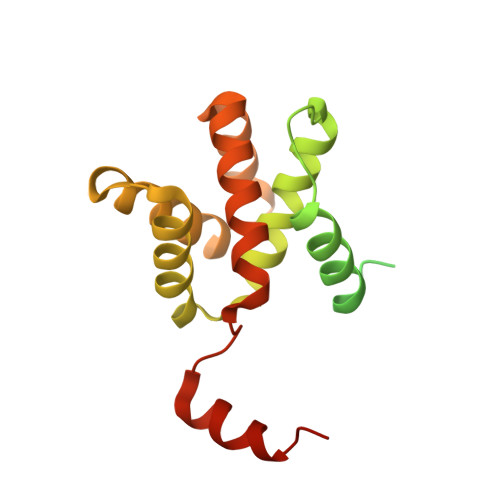
Structure of the Reston ebolavirus VP30 C-terminal domain.

Clifton, M.C.Kirchdoerfer, R.N.Atkins, K.Abendroth, J.Raymond, A.Grice, R.Barnes, S.Moen, S.Lorimer, D.Edwards, T.E.Myler, P.J.Saphire, E.O.

(2014) Acta Crystallogr Sect F Struct Biol Cryst Commun 70: 457-460

  • DOI: https://doi.org/10.1107/S2053230X14003811
  • Primary Citation Related Structures: 
    3V7O

  • PubMed Abstract: 

    The ebolaviruses can cause severe hemorrhagic fever. Essential to the ebolavirus life cycle is the protein VP30, which serves as a transcriptional cofactor. Here, the crystal structure of the C-terminal, NP-binding domain of VP30 from Reston ebolavirus is presented. Reston VP30 and Ebola VP30 both form homodimers, but the dimeric interfaces are rotated relative to each other, suggesting subtle inherent differences or flexibility in the dimeric interface.


  • Organizational Affiliation
    • Seattle Structural Genomics Center for Infectious Disease (SSGCID), 307 Westlake Avenue North, Suite 500, Seattle, WA 98109, USA.

Macromolecule Content 

  • Total Structure Weight: 51.69 kDa 
  • Atom Count: 2,715 
  • Modeled Residue Count: 329 
  • Deposited Residue Count: 454 
  • Unique protein chains: 1

Macromolecules

Find similar proteins by:|  3D Structure
Entity ID: 1
MoleculeChains  Sequence LengthOrganismDetailsImage
Minor nucleoprotein VP30
A, B
227Reston ebolavirus - Reston (1989)Mutation(s): 0 
Gene Names: VP30
UniProt
Find proteins for Q8JPX6 (Reston ebolavirus (strain Reston-89))
Explore Q8JPX6 
Go to UniProtKB:  Q8JPX6
Entity Groups
Sequence Clusters30% Identity50% Identity70% Identity90% Identity95% Identity100% Identity
UniProt GroupQ8JPX6
Sequence Annotations
Expand
Reference Sequence

Experimental Data & Validation

Experimental Data

  • Method: X-RAY DIFFRACTION
  • Resolution: 2.25 Å
  • R-Value Free:  0.226 (Depositor), 0.228 (DCC) 
  • R-Value Work:  0.193 (Depositor), 0.196 (DCC) 
  • R-Value Observed: 0.195 (Depositor) 
Space Group: P 21 21 21
Unit Cell:
Length ( Å )Angle ( ˚ )
a = 49.31α = 90
b = 93.74β = 90
c = 111.22γ = 90
Software Package:
Software NamePurpose
XSCALEdata scaling
PHASERphasing
REFMACrefinement
PDB_EXTRACTdata extraction
XDSdata reduction

Structure Validation

View Full Validation Report


Entry History 

Deposition Data

Revision History  (Full details and data files)

  • Version 1.0: 2012-02-08
    Type: Initial release
  • Version 1.1: 2014-05-14
    Changes: Database references
  • Version 1.2: 2023-09-13
    Changes: Data collection, Database references, Derived calculations, Refinement description
  • Version 1.3: 2025-10-22
    Changes: Structure summary