3V42 | pdb_00003v42

Crystal structure of renal tumor suppressor protein, folliculin


Experimental Data Snapshot

  • Method: X-RAY DIFFRACTION
  • Resolution: 2.00 Å
  • R-Value Free: 
    0.268 (Depositor), 0.275 (DCC) 
  • R-Value Work: 
    0.204 (Depositor), 0.215 (DCC) 
  • R-Value Observed: 
    0.207 (Depositor) 

Starting Model: experimental
View more details

wwPDB Validation 3D Report Full Report

Validation slider image for 3V42

This is version 1.3 of the entry. See complete history

Literature

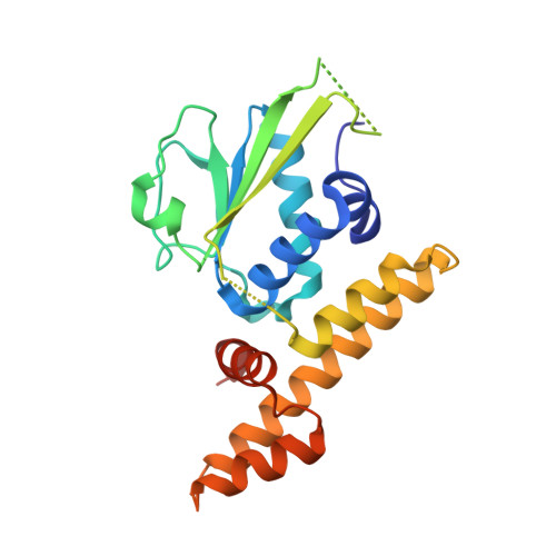
Crystal structure of folliculin reveals a hidDENN function in genetically inherited renal cancer.

Nookala, R.K.Langemeyer, L.Pacitto, A.Ochoa-Montano, B.Donaldson, J.C.Blaszczyk, B.K.Chirgadze, D.Y.Barr, F.A.Bazan, J.F.Blundell, T.L.

(2012) Open Biol 2: 120071-120071

  • DOI: https://doi.org/10.1098/rsob.120071
  • Primary Citation Related Structures: 
    3V42

  • PubMed Abstract: 

    Mutations in the renal tumour suppressor protein, folliculin, lead to proliferative skin lesions, lung complications and renal cell carcinoma. Folliculin has been reported to interact with AMP-activated kinase, a key component of the mammalian target of rapamycin pathway. Most cancer-causing mutations lead to a carboxy-terminal truncation of folliculin, pointing to a functional importance of this domain in tumour suppression. We present here the crystal structure of folliculin carboxy-terminal domain and demonstrate that it is distantly related to differentially expressed in normal cells and neoplasia (DENN) domain proteins, a family of Rab guanine nucleotide exchange factors (GEFs). Using biochemical analysis, we show that folliculin has GEF activity, indicating that folliculin is probably a distantly related member of this class of Rab GEFs.


  • Organizational Affiliation
    • Department of Biochemistry , University of Cambridge, Sanger Building, 80 Tennis Court Road, Cambridge CB2 1GA, UK. rn229@cam.ac.uk

Macromolecule Content 

  • Total Structure Weight: 50.93 kDa 
  • Atom Count: 3,308 
  • Modeled Residue Count: 395 
  • Deposited Residue Count: 452 
  • Unique protein chains: 1

Macromolecules

Find similar proteins by:|  3D Structure
Entity ID: 1
MoleculeChains  Sequence LengthOrganismDetailsImage
Folliculin
A, B
226Homo sapiensMutation(s): 3 
Gene Names: BHDFLCN
UniProt & NIH Common Fund Data Resources
Find proteins for Q8NFG4 (Homo sapiens)
Explore Q8NFG4 
Go to UniProtKB:  Q8NFG4
PHAROS:  Q8NFG4
GTEx:  ENSG00000154803 
Entity Groups
Sequence Clusters30% Identity50% Identity70% Identity90% Identity95% Identity100% Identity
UniProt GroupQ8NFG4
Sequence Annotations
Expand
Reference Sequence

Experimental Data & Validation

Experimental Data

  • Method: X-RAY DIFFRACTION
  • Resolution: 2.00 Å
  • R-Value Free:  0.268 (Depositor), 0.275 (DCC) 
  • R-Value Work:  0.204 (Depositor), 0.215 (DCC) 
  • R-Value Observed: 0.207 (Depositor) 
Space Group: C 2 2 21
Unit Cell:
Length ( Å )Angle ( ˚ )
a = 86.05α = 90
b = 99.951β = 90
c = 107.584γ = 90
Software Package:
Software NamePurpose
DNAdata collection
PHENIXmodel building
REFMACrefinement
XDSdata reduction
SCALAdata scaling
PHENIXphasing

Structure Validation

View Full Validation Report


Entry History 

Deposition Data

Revision History  (Full details and data files)

  • Version 1.0: 2012-08-15
    Type: Initial release
  • Version 1.1: 2012-09-26
    Changes: Database references
  • Version 1.2: 2024-02-28
    Changes: Data collection, Database references
  • Version 1.3: 2024-04-03
    Changes: Refinement description