3V2N | pdb_00003v2n

COMPcc in complex with fatty acids


Experimental Data Snapshot

  • Method: X-RAY DIFFRACTION
  • Resolution: 1.80 Å
  • R-Value Free: 
    0.275 (Depositor), 0.275 (DCC) 
  • R-Value Work: 
    0.211 (Depositor), 0.211 (DCC) 

Starting Model: experimental
View more details

wwPDB Validation 3D Report Full Report

Validation slider image for 3V2N

Ligand Structure Quality Assessment 


This is version 1.3 of the entry. See complete history

Literature

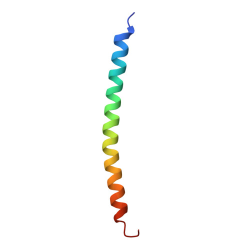
The pentameric channel of COMPcc in complex with different fatty acids.

MacFarlane, A.A.Orriss, G.Okun, N.Meier, M.Klonisch, T.Khajehpour, M.Stetefeld, J.

(2012) PLoS One 7: e48130-e48130

  • DOI: https://doi.org/10.1371/journal.pone.0048130
  • Primary Citation Related Structures: 
    3V2N, 3V2P, 3V2Q

  • PubMed Abstract: 

    COMPcc forms a pentameric left-handed coiled coil that is known to bind hydrophilic signaling molecules such as vitamin D(3), and vitamin A. In an integrated approach we reveal the unique binding properties of COMPcc for saturated and unsaturated fatty acids. Our observations suggest that residues Met33 (gating pore), Thr40/Asn41 (water chamber) and Gln54 (electrostatic trap) are key elements for the binding of fatty acids by COMPcc. In addition, this work characterizes the binding of various fatty acids to COMPcc using fluorescence spectroscopy. Our findings reveal a binding trend within the hydrophobic channel of COMPcc, namely, that is driven by length of the methylene tail and incorporation of unsaturation. The unique binding properties imply that COMPcc may be involved in signalling functions in which hydrophilic ligands are involved. The pentameric channel is a unique carrier for lipophilic compounds. This opens the exciting possibility that COMPcc could be developed as a targeted drug delivery system.


  • Organizational Affiliation
    • Department of Chemistry, University of Manitoba, Canada.

Macromolecule Content 

  • Total Structure Weight: 26.44 kDa 
  • Atom Count: 2,178 
  • Modeled Residue Count: 225 
  • Deposited Residue Count: 225 
  • Unique protein chains: 1

Macromolecules

Find similar proteins by:|  3D Structure
Entity ID: 1
MoleculeChains  Sequence LengthOrganismDetailsImage
Cartilage Oligomerization matrix protein (coiled-coil domain)
A, B, C, D, E
45Mus musculusMutation(s): 0 
UniProt
Find proteins for Q9R0G6 (Mus musculus)
Explore Q9R0G6 
Go to UniProtKB:  Q9R0G6
Entity Groups
Sequence Clusters30% Identity50% Identity70% Identity90% Identity95% Identity100% Identity
UniProt GroupQ9R0G6
Sequence Annotations
Expand
Reference Sequence

Small Molecules

Ligands 1 Unique
IDChains Name / Formula / InChI Key2D Diagram3D Interactions
MYR

Query on MYR


Download:Ideal Coordinates CCD File
F [auth A]MYRISTIC ACID
C14 H28 O2
TUNFSRHWOTWDNC-UHFFFAOYSA-N

Experimental Data & Validation

Experimental Data

  • Method: X-RAY DIFFRACTION
  • Resolution: 1.80 Å
  • R-Value Free:  0.275 (Depositor), 0.275 (DCC) 
  • R-Value Work:  0.211 (Depositor), 0.211 (DCC) 
Space Group: P 1 21 1
Unit Cell:
Length ( Å )Angle ( ˚ )
a = 38.356α = 90
b = 49.391β = 103.87
c = 54.82γ = 90
Software Package:
Software NamePurpose
CNSrefinement
PDB_EXTRACTdata extraction
HKL-2000data collection
DENZOdata reduction
SCALAdata scaling
AMoREphasing

Structure Validation

View Full Validation Report


Ligand Structure Quality Assessment 


Entry History 

Deposition Data

Revision History  (Full details and data files)

  • Version 1.0: 2013-01-16
    Type: Initial release
  • Version 1.1: 2013-03-20
    Changes: Database references
  • Version 1.2: 2023-09-13
    Changes: Data collection, Database references, Derived calculations, Refinement description
  • Version 1.3: 2024-10-30
    Changes: Structure summary