3UWN | pdb_00003uwn

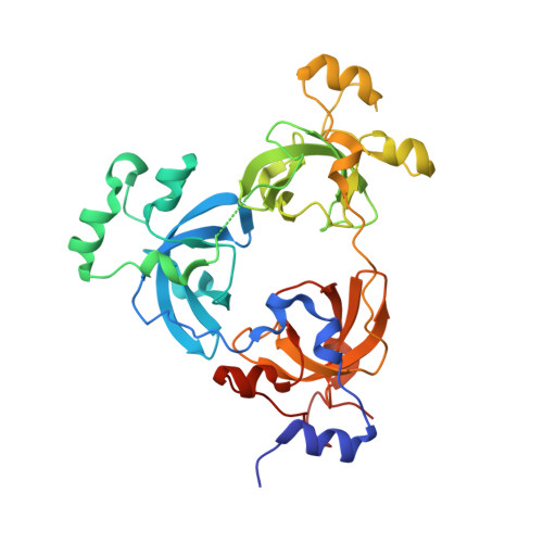
The 3-MBT repeat domain of L3MBTL1 in complex with a methyl-lysine mimic


Experimental Data Snapshot

  • Method: X-RAY DIFFRACTION
  • Resolution: 2.15 Å
  • R-Value Free: 
    0.267 (Depositor), 0.259 (DCC) 
  • R-Value Work: 
    0.231 (Depositor), 0.233 (DCC) 
  • R-Value Observed: 
    0.233 (Depositor) 

Starting Model: experimental
View more details

wwPDB Validation 3D Report Full Report

Validation slider image for 3UWN

Ligand Structure Quality Assessment 


This is version 1.1 of the entry. See complete history

Literature

Macromolecule Content 

  • Total Structure Weight: 38.28 kDa 
  • Atom Count: 2,710 
  • Modeled Residue Count: 318 
  • Deposited Residue Count: 331 
  • Unique protein chains: 1

Macromolecules

Find similar proteins by:|  3D Structure
Entity ID: 1
MoleculeChains  Sequence LengthOrganismDetailsImage
Lethal(3)malignant brain tumor-like protein 1331Homo sapiensMutation(s): 0 
Gene Names: L3MBTL1KIAA0681L3MBTL3MBTL
UniProt & NIH Common Fund Data Resources
Find proteins for Q9Y468 (Homo sapiens)
Explore Q9Y468 
Go to UniProtKB:  Q9Y468
PHAROS:  Q9Y468
GTEx:  ENSG00000185513 
Entity Groups
Sequence Clusters30% Identity50% Identity70% Identity90% Identity95% Identity100% Identity
UniProt GroupQ9Y468
Sequence Annotations
Expand
Reference Sequence

Small Molecules

Ligands 2 Unique
IDChains Name / Formula / InChI Key2D Diagram3D Interactions
UWN

Query on UWN


Download:Ideal Coordinates CCD File
B [auth A][2-(phenylamino)benzene-1,4-diyl]bis{[4-(pyrrolidin-1-yl)piperidin-1-yl]methanone}
C32 H43 N5 O2
PQOOIERVZAXHBP-UHFFFAOYSA-N
UNX

Query on UNX


Download:Ideal Coordinates CCD File
C [auth A]
D [auth A]
E [auth A]
F [auth A]
G [auth A]
C [auth A],
D [auth A],
E [auth A],
F [auth A],
G [auth A],
H [auth A],
I [auth A],
J [auth A],
K [auth A],
L [auth A],
M [auth A],
N [auth A],
O [auth A],
P [auth A]
UNKNOWN ATOM OR ION
X
Binding Affinity Annotations 
IDSourceBinding Affinity
UWN BindingDB:  3UWN Kd: 9400 (nM) from 1 assay(s)
IC50: min: 2000, max: 8900 (nM) from 3 assay(s)

Experimental Data & Validation

Experimental Data

  • Method: X-RAY DIFFRACTION
  • Resolution: 2.15 Å
  • R-Value Free:  0.267 (Depositor), 0.259 (DCC) 
  • R-Value Work:  0.231 (Depositor), 0.233 (DCC) 
  • R-Value Observed: 0.233 (Depositor) 
Space Group: P 21 21 2
Unit Cell:
Length ( Å )Angle ( ˚ )
a = 85.774α = 90
b = 94.53β = 90
c = 58.955γ = 90
Software Package:
Software NamePurpose
SCALAdata scaling
PHASERphasing
REFMACrefinement
PDB_EXTRACTdata extraction
XDSdata reduction

Structure Validation

View Full Validation Report


Ligand Structure Quality Assessment 


Entry History 

Revision History  (Full details and data files)

  • Version 1.0: 2012-03-07
    Type: Initial release
  • Version 1.1: 2023-09-13
    Changes: Data collection, Database references, Derived calculations, Refinement description