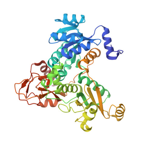
Crystal structure of Brucella abortus deoxyxylulose-5-phosphate reductoisomerase-like (DRL) enzyme involved in isoprenoid biosynthesis.
Perez-Gil, J., Calisto, B.M., Behrendt, C., Kurz, T., Fita, I., Rodriguez-Concepcion, M.(2012) J Biol Chem 287: 15803-15809
- PubMed: 22442144 Search on PubMedSearch on PubMed Central
- DOI: https://doi.org/10.1074/jbc.M112.354811
- Primary Citation Related Structures:
3UPL, 3UPY - PubMed Abstract:
Most bacteria use the 2-C-methyl-D-erythritol 4-phosphate (MEP) pathway for the synthesis of their essential isoprenoid precursors. The absence of the MEP pathway in humans makes it a promising new target for the development of much needed new and safe antimicrobial drugs. However, bacteria show a remarkable metabolic plasticity for isoprenoid production. For example, the NADPH-dependent production of MEP from 1-deoxy-D-xylulose 5-phosphate in the first committed step of the MEP pathway is catalyzed by 1-deoxy-D-xylulose-5-phosphate reductoisomerase (DXR) in most bacteria, whereas an unrelated DXR-like (DRL) protein was recently found to catalyze the same reaction in some organisms, including the emerging human and animal pathogens Bartonella and Brucella. Here, we report the x-ray crystal structures of the Brucella abortus DRL enzyme in its apo form and in complex with the broad-spectrum antibiotic fosmidomycin solved to 1.5 and 1.8 Å resolution, respectively. DRL is a dimer, with each polypeptide folding into three distinct domains starting with the NADPH-binding domain, in resemblance to the structure of bacterial DXR enzymes. Other than that, DRL and DXR show a low structural relationship, with a different disposition of the domains and a topologically unrelated C-terminal domain. In particular, the active site of DRL presents a unique arrangement, suggesting that the design of drugs that would selectively inhibit DRL-harboring pathogens without affecting beneficial or innocuous bacteria harboring DXR should be feasible. As a proof of concept, we identified two strong DXR inhibitors that have virtually no effect on DRL activity.
- Department of Molecular Genetics, Centre for Research in Agricultural Genomics (CRAG), Consejo Superior de Investigaciones Científicas (CSIC)-Institut de Recerca i Tecnologia Agroalimentàries (IRTA)-Universitat Autònoma de Barcelona (UAB), 08193 Barcelona, Spain.
Organizational Affiliation: