3UOB | pdb_00003uob

Crystal structure of Human Thymine DNA Glycosylase Bound to Substrate Analog 2'-deoxy-2'-beta-fluoro-cytidine


Experimental Data Snapshot

  • Method: X-RAY DIFFRACTION
  • Resolution: 3.01 Å
  • R-Value Free: 
    0.276 (Depositor), 0.243 (DCC) 
  • R-Value Work: 
    0.219 (Depositor), 0.234 (DCC) 
  • R-Value Observed: 
    0.225 (Depositor) 

Starting Model: experimental
View more details

wwPDB Validation 3D Report Full Report

Validation slider image for 3UOB

This is version 1.2 of the entry. See complete history

Literature

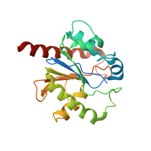
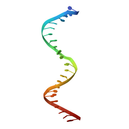
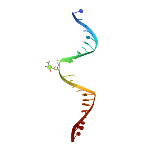
Thymine DNA glycosylase specifically recognizes 5-carboxylcytosine-modified DNA.

Zhang, L.Lu, X.Lu, J.Liang, H.Dai, Q.Xu, G.L.Luo, C.Jiang, H.He, C.

(2012) Nat Chem Biol 8: 328-330

  • DOI: https://doi.org/10.1038/nchembio.914
  • Primary Citation Related Structures: 
    3UO7, 3UOB

  • PubMed Abstract: 

    Human thymine DNA glycosylase (hTDG) efficiently excises 5-carboxylcytosine (5caC), a key oxidation product of 5-methylcytosine in genomic DNA, in a recently discovered cytosine demethylation pathway. We present here the crystal structures of the hTDG catalytic domain in complex with duplex DNA containing either 5caC or a fluorinated analog. These structures, together with biochemical and computational analyses, reveal that 5caC is specifically recognized in the active site of hTDG, supporting the role of TDG in mammalian 5-methylcytosine demethylation.


  • Organizational Affiliation
    • Department of Chemistry and Institute for Biophysical Dynamics, The University of Chicago, Chicago, Illinois, USA.

Macromolecule Content 

  • Total Structure Weight: 59.73 kDa 
  • Atom Count: 3,764 
  • Modeled Residue Count: 399 
  • Deposited Residue Count: 448 
  • Unique protein chains: 1
  • Unique nucleic acid chains: 2

Macromolecules


Find similar proteins by:|  3D Structure
Entity ID: 3
MoleculeChains  Sequence LengthOrganismDetailsImage
G/T mismatch-specific thymine DNA glycosylaseC [auth A],
D [auth B]
201Homo sapiensMutation(s): 0 
Gene Names: TDG
EC: 3.2.2.29
UniProt & NIH Common Fund Data Resources
Find proteins for Q13569 (Homo sapiens)
Explore Q13569 
Go to UniProtKB:  Q13569
PHAROS:  Q13569
GTEx:  ENSG00000139372 
Entity Groups
Sequence Clusters30% Identity50% Identity70% Identity90% Identity95% Identity100% Identity
UniProt GroupQ13569
Sequence Annotations
Expand
Reference Sequence
Find similar nucleic acids by:  Sequence
Entity ID: 1
MoleculeChains LengthOrganismImage
5'-D(*CP*AP*GP*CP*TP*CP*TP*GP*TP*AP*CP*GP*TP*GP*AP*GP*CP*AP*GP*TP*GP*GP*A)-3'A [auth C]23N/A
Sequence Annotations
Expand
Reference Sequence
Find similar nucleic acids by:  Sequence
Entity ID: 2
MoleculeChains LengthOrganismImage
5'-D(*CP*CP*AP*CP*TP*GP*CP*TP*CP*AP*(1FC)P*GP*TP*AP*CP*AP*GP*AP*GP*CP*TP*GP*T)-3'B [auth D]23N/A
Sequence Annotations
Expand
Reference Sequence

Experimental Data & Validation

Experimental Data

  • Method: X-RAY DIFFRACTION
  • Resolution: 3.01 Å
  • R-Value Free:  0.276 (Depositor), 0.243 (DCC) 
  • R-Value Work:  0.219 (Depositor), 0.234 (DCC) 
  • R-Value Observed: 0.225 (Depositor) 
Space Group: P 65
Unit Cell:
Length ( Å )Angle ( ˚ )
a = 163.418α = 90
b = 163.418β = 90
c = 54.701γ = 120
Software Package:
Software NamePurpose
HKL-2000data collection
PHASESphasing
PHENIXrefinement
HKL-3000data reduction
HKL-3000data scaling

Structure Validation

View Full Validation Report


Entry History 

Deposition Data

Revision History  (Full details and data files)

  • Version 1.0: 2012-02-15
    Type: Initial release
  • Version 1.1: 2015-06-03
    Changes: Database references
  • Version 1.2: 2023-09-13
    Changes: Data collection, Database references, Derived calculations, Refinement description