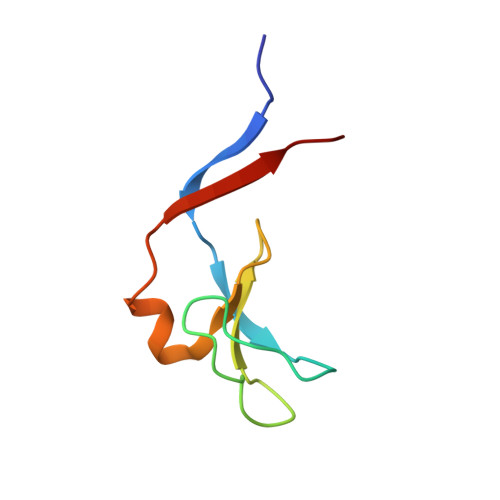
Structural and Functional Characterization of an Anesthetic Binding Site in the Second Cysteine-Rich Domain of Protein Kinase Cdelta
Shanmugasundararaj, S., Das, J., Sandberg, W.S., Zhou, X., Wang, D., Messing, R.O., Bruzik, K.S., Stehle, T., Miller, K.W.(2012) Biophys J 103: 2331-2340
- PubMed: 23283232
- DOI: https://doi.org/10.1016/j.bpj.2012.10.034
- Primary Citation of Related Structures:
3UEJ, 3UEY, 3UFF, 3UGD, 3UGI, 3UGL - PubMed Abstract:
Elucidating the principles governing anesthetic-protein interactions requires structural determinations at high resolutions not yet achieved with ion channels. Protein kinase C (PKC) activity is modulated by general anesthetics. We solved the structure of the phorbol-binding domain (C1B) of PKCδ complexed with an ether (methoxymethylcycloprane) and with an alcohol (cyclopropylmethanol) at 1.36-Å resolution. The cyclopropane rings of both agents displace a single water molecule in a surface pocket adjacent to the phorbol-binding site, making van der Waals contacts with the backbone and/or side chains of residues Asn-237 to Ser-240. Surprisingly, two water molecules anchored in a hydrogen-bonded chain between Thr-242 and Lys-260 impart elasticity to one side of the binding pocket. The cyclopropane ring takes part in π-acceptor hydrogen bonds with the amide of Met-239. There is a crucial hydrogen bond between the oxygen atoms of the anesthetics and the hydroxyl of Tyr-236. A Tyr-236-Phe mutation results in loss of binding. Thus, both van der Waals interactions and hydrogen-bonding are essential for binding to occur. Ethanol failed to bind because it is too short to benefit from both interactions. Cyclopropylmethanol inhibited phorbol-ester-induced PKCδ activity, but failed to do so in PKCδ containing the Tyr-236-Phe mutation.
- Department of Anesthesia and Critical Care, Massachusetts General Hospital, Boston, MA, USA.
Organizational Affiliation:


