3UC7 | pdb_00003uc7

Trp-cage cyclo-TC1 - monoclinic crystal form


Experimental Data Snapshot

  • Method: X-RAY DIFFRACTION
  • Resolution: 1.10 Å
  • R-Value Free: 
    0.194 (Depositor), 0.215 (DCC) 
  • R-Value Work: 
    0.149 (Depositor) 
  • R-Value Observed: 
    0.151 (Depositor) 

Starting Model: experimental
View more details

wwPDB Validation 3D Report Full Report

Validation slider image for 3UC7

This is version 1.5 of the entry. See complete history

Literature

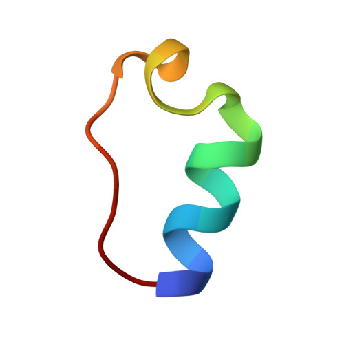
Crystal and NMR structures of a Trp-cage mini-protein benchmark for computational fold prediction.

Scian, M.Lin, J.C.Le Trong, I.Makhatadze, G.I.Stenkamp, R.E.Andersen, N.H.

(2012) Proc Natl Acad Sci U S A 109: 12521-12525

  • DOI: https://doi.org/10.1073/pnas.1121421109
  • Primary Citation Related Structures: 
    2LL5, 3UC7, 3UC8

  • PubMed Abstract: 

    To provide high-resolution X-ray crystallographic structures of a peptide with the Trp-cage fold, we prepared a cyclized version of this motif. Cyclized Trp-cage is remarkably stable and afforded two crystal forms suitable for X-ray diffraction. The resulting higher resolution crystal structures validate the prior NMR models and provide explanations for experimental observations that could not be rationalized by NMR structural data, including the structural basis for the increase in fold stability associated with motif cyclization and the manner in which a polar serine side chain is accommodated in the hydrophobic interior. A hexameric oligomer of the cyclic peptide is found in both crystal forms and indicates that under appropriate conditions, this minimized system may also serve as a model for protein-protein interactions.


  • Organizational Affiliation
    • Department of Chemistry, University of Washington, Seattle, WA 98195, USA. scian@u.washington.edu

Macromolecule Content 

  • Total Structure Weight: 12.94 kDa 
  • Atom Count: 1,049 
  • Modeled Residue Count: 132 
  • Deposited Residue Count: 132 
  • Unique protein chains: 1

Macromolecules

Find similar proteins by:  Sequence   |   3D Structure  
Entity ID: 1
MoleculeChains  Sequence LengthOrganismDetailsImage
Cyclo-TC1
A, B, C, D, E
A, B, C, D, E, F
22N/AMutation(s): 0 

Experimental Data & Validation

Experimental Data

  • Method: X-RAY DIFFRACTION
  • Resolution: 1.10 Å
  • R-Value Free:  0.194 (Depositor), 0.215 (DCC) 
  • R-Value Work:  0.149 (Depositor) 
  • R-Value Observed: 0.151 (Depositor) 
Space Group: C 1 2 1
Unit Cell:
Length ( Å )Angle ( ˚ )
a = 57.697α = 90
b = 48.748β = 116.91
c = 35.259γ = 90
Software Package:
Software NamePurpose
DENZOdata reduction
SCALEPACKdata scaling
PHASERphasing
REFMACrefinement
PDB_EXTRACTdata extraction
Blu-Icedata collection

Structure Validation

View Full Validation Report


Entry History 

Deposition Data

Revision History  (Full details and data files)

  • Version 1.0: 2012-07-18
    Type: Initial release
  • Version 1.1: 2012-08-01
    Changes: Database references
  • Version 1.2: 2012-08-15
    Changes: Database references
  • Version 1.3: 2017-11-08
    Changes: Refinement description
  • Version 1.4: 2023-09-13
    Changes: Data collection, Database references, Derived calculations, Refinement description
  • Version 1.5: 2024-10-30
    Changes: Structure summary