3UBU | pdb_00003ubu

Crystal structure of agkisacucetin, a GpIb-binding snaclec (snake C-type lectin) that inhibits platelet


Experimental Data Snapshot

  • Method: X-RAY DIFFRACTION
  • Resolution: 1.91 Å
  • R-Value Free: 
    0.222 (Depositor), 0.307 (DCC) 
  • R-Value Work: 
    0.192 (Depositor) 
  • R-Value Observed: 
    0.193 (Depositor) 

wwPDB Validation 3D Report Full Report

Validation slider image for 3UBU

This is version 1.1 of the entry. See complete history

Literature

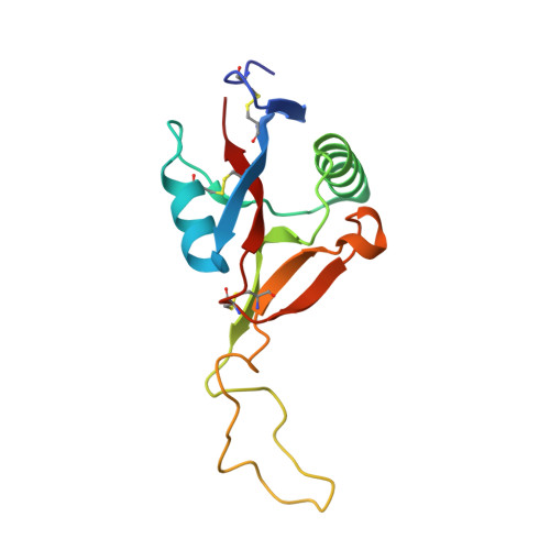
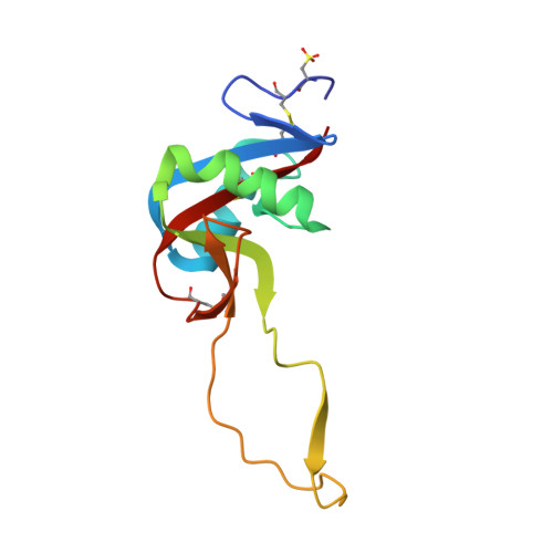
Crystal structure of agkisacucetin, a Gpib-binding snake C-type lectin that inhibits platelet adhesion and aggregation.

Gao, Y.Ge, H.Chen, H.Li, H.Liu, Y.Chen, L.Li, X.Liu, J.Niu, L.Teng, M.

(2012) Proteins 

  • DOI: https://doi.org/10.1002/prot.24060
  • Primary Citation Related Structures: 
    3UBU

  • PubMed Abstract: 

    Agkisacucetin is a snake C-type lectin isolated from the venom of Agkistrodon acutus (A. acutus). It binds specifically to the platelet glycoprotein (GP) Ib and prevents the von Willebrand factor (VWF) accessing it. We determined the crystal structure of agkisacucetin to 1.9Å resolution. The structure of agkisacucetin has an (αβ) fold similar to another GPIb-binding protein, flavocetin-A, but lacks the C-terminal cysteine in the β-subunit, does not form (βα)(4) tetramers, and does not cluster GPIbs, like flavocetin-A.


  • Organizational Affiliation
    • Hefei National Laboratory for Physical Sciences at Microscale, School of Life Sciences, University of Science and Technology of China, Hefei, Anhui 230026, China.

Macromolecule Content 

  • Total Structure Weight: 30.16 kDa 
  • Atom Count: 2,239 
  • Modeled Residue Count: 256 
  • Deposited Residue Count: 257 
  • Unique protein chains: 2

Macromolecules

Find similar proteins by:|  3D Structure
Entity ID: 1
MoleculeChains  Sequence LengthOrganismDetailsImage
Agglucetin subunit alpha-1131Deinagkistrodon acutusMutation(s): 0 
UniProt
Find proteins for Q8JIV9 (Deinagkistrodon acutus)
Explore Q8JIV9 
Go to UniProtKB:  Q8JIV9
Entity Groups
Sequence Clusters30% Identity50% Identity70% Identity90% Identity95% Identity100% Identity
UniProt GroupQ8JIV9
Sequence Annotations
Expand
Reference Sequence
Find similar proteins by:|  3D Structure
Entity ID: 2
MoleculeChains  Sequence LengthOrganismDetailsImage
Agglucetin subunit beta-2126Deinagkistrodon acutusMutation(s): 0 
UniProt
Find proteins for Q8AYA3 (Deinagkistrodon acutus)
Explore Q8AYA3 
Go to UniProtKB:  Q8AYA3
Entity Groups
Sequence Clusters30% Identity50% Identity70% Identity90% Identity95% Identity100% Identity
UniProt GroupQ8AYA3
Sequence Annotations
Expand
Reference Sequence

Experimental Data & Validation

Experimental Data

  • Method: X-RAY DIFFRACTION
  • Resolution: 1.91 Å
  • R-Value Free:  0.222 (Depositor), 0.307 (DCC) 
  • R-Value Work:  0.192 (Depositor) 
  • R-Value Observed: 0.193 (Depositor) 
Space Group: P 21 21 21
Unit Cell:
Length ( Å )Angle ( ˚ )
a = 30.485α = 90
b = 58.052β = 90
c = 141.537γ = 90
Software Package:
Software NamePurpose
HKL-2000data collection
MOLREPphasing
REFMACrefinement
HKL-2000data reduction
HKL-2000data scaling

Structure Validation

View Full Validation Report


Entry History 

Deposition Data

Revision History  (Full details and data files)

  • Version 1.0: 2012-04-11
    Type: Initial release
  • Version 1.1: 2025-03-26
    Changes: Data collection, Database references, Derived calculations, Structure summary