


Crystal structure of a chitin binding domain from Burkholderia pseudomallei
Seattle Structural Genomics Center for Infectious Disease (SSGCID), Fox III, D., Gardberg, A., Armour, B., Staker, B., Stewart, L.To be published.
Experimental Data Snapshot
Starting Model: experimental
View more details
wwPDB Validation 3D Report Full Report

Macromolecule Content
Entity ID: 1 | |||||
|---|---|---|---|---|---|
| Molecule | Chains | Sequence Length | Organism | Details | Image |
| Chitin binding domain | 216 | Burkholderia pseudomallei 1710b | Mutation(s): 0 Gene Names: BURPS1710b_0114 | 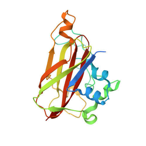 | |
UniProt | |||||
Entity Groups | |||||
| Sequence Clusters | 30% Identity50% Identity70% Identity90% Identity95% Identity100% Identity | ||||
| UniProt Group | Q3JY22 | ||||
Sequence AnnotationsExpand | |||||
Reference Sequence | |||||
| Ligands 2 Unique | |||||
|---|---|---|---|---|---|
| ID | Chains | Name / Formula / InChI Key | 2D Diagram | 3D Interactions | |
| GOL Download:Ideal Coordinates CCD File | I [auth A], L [auth D], N [auth F] | GLYCEROL C3 H8 O3 PEDCQBHIVMGVHV-UHFFFAOYSA-N |  | ||
| NO3 Download:Ideal Coordinates CCD File | G [auth A], H [auth A], J [auth B], K [auth C], M [auth E] | NITRATE ION N O3 NHNBFGGVMKEFGY-UHFFFAOYSA-N |  | ||
| Length ( Å ) | Angle ( ˚ ) |
|---|---|
| a = 70.92 | α = 120.04 |
| b = 74.78 | β = 98.04 |
| c = 74.77 | γ = 102.98 |
| Software Name | Purpose |
|---|---|
| XSCALE | data scaling |
| PHASER | phasing |
| REFMAC | refinement |
| PDB_EXTRACT | data extraction |


