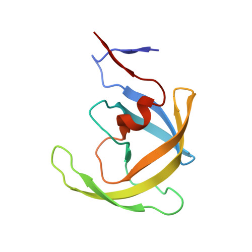
3U7S

HIV PR drug resistant patient's variant in complex with darunavir


Experimental Data Snapshot

  • Method: X-RAY DIFFRACTION
  • Resolution: 2.05 Å
  • R-Value Free: 0.268 
  • R-Value Work: 0.190 
  • R-Value Observed: 0.194 

wwPDB Validation   3D Report Full Report


Ligand Structure Quality Assessment 


This is version 1.2 of the entry. See complete history


Literature

Molecular characterization of clinical isolates of human immunodeficiency virus resistant to the protease inhibitor darunavir.

Saskova, K.G.Kozisek, M.Rezacova, P.Brynda, J.Yashina, T.Kagan, R.M.Konvalinka, J.

(2009) J Virol 83: 8810-8818

  • DOI: https://doi.org/10.1128/JVI.00451-09
  • Primary Citation of Related Structures:  
    3GGU, 3U7S

  • PubMed Abstract: 

    Darunavir is the most recently approved human immunodeficiency virus (HIV) protease (PR) inhibitor (PI) and is active against many HIV type 1 PR variants resistant to earlier-generation PIs. Darunavir shows a high genetic barrier to resistance development, and virus strains with lower sensitivity to darunavir have a higher number of PI resistance-associated mutations than viruses resistant to other PIs. In this work, we have enzymologically and structurally characterized a number of highly mutated clinically derived PRs with high levels of phenotypic resistance to darunavir. With 18 to 21 amino acid residue changes, the PR variants studied in this work are the most highly mutated HIV PR species ever studied by means of enzyme kinetics and X-ray crystallography. The recombinant proteins showed major defects in substrate binding, while the substrate turnover was less affected. Remarkably, the overall catalytic efficiency of the recombinant PRs (5% that of the wild-type enzyme) is still sufficient to support polyprotein processing and particle maturation in the corresponding viruses. The X-ray structures of drug-resistant PRs complexed with darunavir suggest that the impaired inhibitor binding could be explained by change in the PR-inhibitor hydrogen bond pattern in the P2' binding pocket due to a substantial shift of the aminophenyl moiety of the inhibitor. Recombinant virus phenotypic characterization, enzyme kinetics, and X-ray structural analysis thus help to explain darunavir resistance development in HIV-positive patients.


  • Organizational Affiliation

    Gilead Sciences and IOCB Research Center, Institute of Organic Chemistry and Biochemistry of the Academy of Sciences of the Czech Republic, v.v.i., Flemingovo n. 2, 166 10 Prague 6, Czech Republic.


Macromolecules
Find similar proteins by:  (by identity cutoff)  |  3D Structure
Entity ID: 1
MoleculeChains Sequence LengthOrganismDetailsImage
Pol polyprotein
A, B
99Human immunodeficiency virus 1Mutation(s): 11 
Gene Names: pol
EC: 3.4.23.16
UniProt
Find proteins for P35963 (Human immunodeficiency virus type 1 group M subtype B (isolate YU-2))
Explore P35963 
Go to UniProtKB:  P35963
Entity Groups  
Sequence Clusters30% Identity50% Identity70% Identity90% Identity95% Identity100% Identity
UniProt GroupP35963
Sequence Annotations
Expand
  • Reference Sequence
Small Molecules
Ligands 2 Unique
IDChains Name / Formula / InChI Key2D Diagram3D Interactions
017
Query on 017

Download Ideal Coordinates CCD File 
C [auth A],
D [auth A]
(3R,3AS,6AR)-HEXAHYDROFURO[2,3-B]FURAN-3-YL(1S,2R)-3-[[(4-AMINOPHENYL)SULFONYL](ISOBUTYL)AMINO]-1-BENZYL-2-HYDROXYPROPYLCARBAMATE
C27 H37 N3 O7 S
CJBJHOAVZSMMDJ-HEXNFIEUSA-N
BME
Query on BME

Download Ideal Coordinates CCD File 
E [auth A],
F [auth B]
BETA-MERCAPTOETHANOL
C2 H6 O S
DGVVWUTYPXICAM-UHFFFAOYSA-N
Experimental Data & Validation

Experimental Data

  • Method: X-RAY DIFFRACTION
  • Resolution: 2.05 Å
  • R-Value Free: 0.268 
  • R-Value Work: 0.190 
  • R-Value Observed: 0.194 
  • Space Group: P 61
Unit Cell:
Length ( Å )Angle ( ˚ )
a = 62.54α = 90
b = 62.54β = 90
c = 82.64γ = 120
Software Package:
Software NamePurpose
MAR345dtbdata collection
REFMACrefinement
MOSFLMdata reduction
SCALAdata scaling
REFMACphasing

Structure Validation

View Full Validation Report


Ligand Structure Quality Assessment 


Entry History 

Deposition Data

Revision History  (Full details and data files)

  • Version 1.0: 2012-01-18
    Type: Initial release
  • Version 1.1: 2018-03-14
    Changes: Advisory, Database references
  • Version 1.2: 2024-02-28
    Changes: Advisory, Data collection, Database references, Derived calculations, Structure summary