3U7F | pdb_00003u7f

Crystal structure of mPNKP catalytic fragment (D170A) bound to single-stranded DNA (TCCTCp)


Experimental Data Snapshot

  • Method: X-RAY DIFFRACTION
  • Resolution: 1.80 Å
  • R-Value Free: 
    0.214 (Depositor), 0.208 (DCC) 
  • R-Value Work: 
    0.169 (Depositor), 0.166 (DCC) 
  • R-Value Observed: 
    0.171 (Depositor) 

wwPDB Validation 3D Report Full Report

Validation slider image for 3U7F

This is version 1.3 of the entry. See complete history

Literature

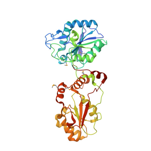
Structural basis for the phosphatase activity of polynucleotide kinase/phosphatase on single- and double-stranded DNA substrates.

Coquelle, N.Havali-Shahriari, Z.Bernstein, N.Green, R.Glover, J.N.

(2011) Proc Natl Acad Sci U S A 108: 21022-21027

  • DOI: https://doi.org/10.1073/pnas.1112036108
  • Primary Citation Related Structures: 
    3U7E, 3U7F, 3U7G, 3U7H

  • PubMed Abstract: 

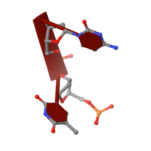
    Polynucleotide kinase/phosphatase (PNKP) is a critical mammalian DNA repair enzyme that generates 5'-phosphate and 3'-hydroxyl groups at damaged DNA termini that are required for subsequent processing by DNA ligases and polymerases. The PNKP phosphatase domain recognizes 3'-phosphate termini within DNA nicks, gaps, or at double- or single-strand breaks. Here we present a mechanistic rationale for the recognition of damaged DNA termini by the PNKP phosphatase domain. The crystal structures of PNKP bound to single-stranded DNA substrates reveals a narrow active site cleft that accommodates a single-stranded substrate in a sequence-independent manner. Biochemical studies suggest that the terminal base pairs of double-stranded substrates near the 3'-phosphate are destabilized by PNKP to allow substrate access to the active site. A positively charged surface distinct from the active site specifically facilitates interactions with double-stranded substrates, providing a complex DNA binding surface that enables the recognition of diverse substrates.


  • Organizational Affiliation
    • Department of Biochemistry, University of Alberta, Edmonton, AB, Canada T6G 2H7.

Macromolecule Content 

  • Total Structure Weight: 44.39 kDa 
  • Atom Count: 3,429 
  • Modeled Residue Count: 381 
  • Deposited Residue Count: 386 
  • Unique protein chains: 1
  • Unique nucleic acid chains: 1

Macromolecules


Find similar proteins by:|  3D Structure
Entity ID: 1
MoleculeChains  Sequence LengthOrganismDetailsImage
Bifunctional polynucleotide phosphatase/kinaseA [auth B]381Mus musculusMutation(s): 1 
Gene Names: Pnkp
EC: 3.1.3.32 (PDB Primary Data), 2.7.1.78 (PDB Primary Data)
UniProt & NIH Common Fund Data Resources
Find proteins for Q9JLV6 (Mus musculus)
Explore Q9JLV6 
Go to UniProtKB:  Q9JLV6
IMPC:  MGI:1891698
Entity Groups
Sequence Clusters30% Identity50% Identity70% Identity90% Identity95% Identity100% Identity
UniProt GroupQ9JLV6
Sequence Annotations
Expand
Reference Sequence
Find similar nucleic acids by:  Sequence
Entity ID: 2
MoleculeChains LengthOrganismImage
DNAB [auth X]5N/A
Sequence Annotations
Expand
Reference Sequence

Small Molecules

Ligands 3 Unique
IDChains Name / Formula / InChI Key2D Diagram3D Interactions
PO4

Query on PO4


Download:Ideal Coordinates CCD File
H [auth B],
I [auth X]
PHOSPHATE ION
O4 P
NBIIXXVUZAFLBC-UHFFFAOYSA-K
GOL

Query on GOL


Download:Ideal Coordinates CCD File
D [auth B],
E [auth B],
F [auth B],
G [auth B]
GLYCEROL
C3 H8 O3
PEDCQBHIVMGVHV-UHFFFAOYSA-N
MG

Query on MG


Download:Ideal Coordinates CCD File
C [auth B]MAGNESIUM ION
Mg
JLVVSXFLKOJNIY-UHFFFAOYSA-N
Modified Residues  2 Unique
IDChains TypeFormula2D DiagramParent
CSO
Query on CSO
A [auth B]L-PEPTIDE LINKINGC3 H7 N O3 SCYS
MSE
Query on MSE
A [auth B]L-PEPTIDE LINKINGC5 H11 N O2 SeMET

Experimental Data & Validation

Experimental Data

  • Method: X-RAY DIFFRACTION
  • Resolution: 1.80 Å
  • R-Value Free:  0.214 (Depositor), 0.208 (DCC) 
  • R-Value Work:  0.169 (Depositor), 0.166 (DCC) 
  • R-Value Observed: 0.171 (Depositor) 
Space Group: P 1 21 1
Unit Cell:
Length ( Å )Angle ( ˚ )
a = 42.6α = 90
b = 62.33β = 91.81
c = 67.97γ = 90
Software Package:
Software NamePurpose
XSCALEdata scaling
PHENIXrefinement
PDB_EXTRACTdata extraction
MxDCdata collection
XDSdata reduction
PHASERphasing

Structure Validation

View Full Validation Report


Entry History 

Deposition Data

Revision History  (Full details and data files)

  • Version 1.0: 2011-12-14
    Type: Initial release
  • Version 1.1: 2012-08-29
    Changes: Database references
  • Version 1.2: 2017-11-08
    Changes: Refinement description
  • Version 1.3: 2024-11-06
    Changes: Data collection, Database references, Derived calculations, Structure summary