3U2A | pdb_00003u2a

Adaptor dependent degradation of a cell-cycle regulator reveals diversity in substrate architectures


Experimental Data Snapshot

  • Method: X-RAY DIFFRACTION
  • Resolution: 1.70 Å
  • R-Value Free: 
    0.206 (Depositor), 0.206 (DCC) 
  • R-Value Work: 
    0.167 (Depositor), 0.166 (DCC) 
  • R-Value Observed: 
    0.169 (Depositor) 

wwPDB Validation 3D Report Full Report

Validation slider image for 3U2A

This is version 1.3 of the entry. See complete history

Literature

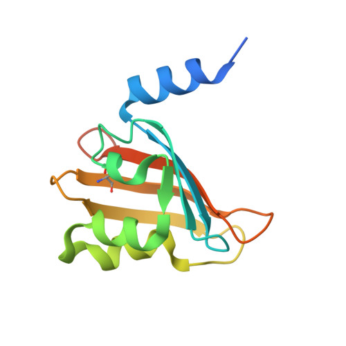
Adaptor-dependent degradation of a cell-cycle regulator uses a unique substrate architecture.

Rood, K.L.Clark, N.E.Stoddard, P.R.Garman, S.C.Chien, P.

(2012) Structure 20: 1223-1232

  • DOI: https://doi.org/10.1016/j.str.2012.04.019
  • Primary Citation Related Structures: 
    3U2A

  • PubMed Abstract: 

    In Caulobacter crescentus, the ClpXP protease degrades several crucial cell-cycle regulators, including the phosphodiesterase PdeA. Degradation of PdeA requires the response regulator CpdR and signals a morphological transition in concert with initiation of DNA replication. Here, we report the structure of a Per-Arnt-Sim (PAS) domain of PdeA and show that it is necessary for CpdR-dependent degradation in vivo and in vitro. CpdR acts as an adaptor, tethering the amino-terminal PAS domain to ClpXP and promoting recognition of the weak carboxyl-terminal degron of PdeA, a combination that ensures processive proteolysis. We identify sites on the PAS domain needed for CpdR recognition and find that one subunit of the PdeA dimer can be delivered to ClpXP by its partner. Finally, we show that improper stabilization of PdeA in vivo alters cellular behavior. These results introduce an adaptor/substrate pair for ClpXP and reveal broad diversity in adaptor-mediated proteolysis.


  • Organizational Affiliation
    • Department of Biochemistry and Molecular Biology, University of Massachusetts, Amherst, Amherst, MA 01003, USA.

Macromolecule Content 

  • Total Structure Weight: 13.98 kDa 
  • Atom Count: 1,037 
  • Modeled Residue Count: 112 
  • Deposited Residue Count: 129 
  • Unique protein chains: 1

Macromolecules

Find similar proteins by:|  3D Structure
Entity ID: 1
MoleculeChains  Sequence LengthOrganismDetailsImage
GGDEF family protein129Caulobacter vibrioidesMutation(s): 0 
Gene Names: CC3396(PdeA)CC_3396
EC: 3.1.4.52
UniProt
Find proteins for A0A0H3CDG5 (Caulobacter vibrioides (strain NA1000 / CB15N))
Explore A0A0H3CDG5 
Go to UniProtKB:  A0A0H3CDG5
Entity Groups
Sequence Clusters30% Identity50% Identity70% Identity90% Identity95% Identity100% Identity
UniProt GroupA0A0H3CDG5
Sequence Annotations
Expand
Reference Sequence

Small Molecules

Modified Residues  1 Unique
IDChains TypeFormula2D DiagramParent
CSO
Query on CSO
A
L-PEPTIDE LINKINGC3 H7 N O3 SCYS

Experimental Data & Validation

Experimental Data

  • Method: X-RAY DIFFRACTION
  • Resolution: 1.70 Å
  • R-Value Free:  0.206 (Depositor), 0.206 (DCC) 
  • R-Value Work:  0.167 (Depositor), 0.166 (DCC) 
  • R-Value Observed: 0.169 (Depositor) 
Space Group: C 1 2 1
Unit Cell:
Length ( Å )Angle ( ˚ )
a = 63.799α = 90
b = 41.895β = 107.52
c = 48.659γ = 90
Software Package:
Software NamePurpose
SCALEPACKdata scaling
MLPHAREphasing
DMphasing
REFMACrefinement
PDB_EXTRACTdata extraction
CrystalCleardata collection
HKL-2000data reduction

Structure Validation

View Full Validation Report


Entry History 

Deposition Data

Revision History  (Full details and data files)

  • Version 1.0: 2012-05-30
    Type: Initial release
  • Version 1.1: 2012-08-29
    Changes: Database references
  • Version 1.2: 2018-04-04
    Changes: Data collection
  • Version 1.3: 2024-11-06
    Changes: Data collection, Database references, Derived calculations, Structure summary