3U16 | pdb_00003u16

Structure of BasE N-terminal domain from Acinetobacter baumannii bound to 6-(p-benzyloxy)phenyl-1-(pyridin-4-ylmethyl)-1H-pyrazolo[3,4-b]pyridine-4-carboxylic acid.


Experimental Data Snapshot

  • Method: X-RAY DIFFRACTION
  • Resolution: 2.10 Å
  • R-Value Free: 
    0.219 (Depositor), 0.220 (DCC) 
  • R-Value Work: 
    0.185 (Depositor) 
  • R-Value Observed: 
    0.187 (Depositor) 

Starting Model: experimental
View more details

wwPDB Validation 3D Report Full Report

Validation slider image for 3U16

Ligand Structure Quality Assessment 


This is version 1.3 of the entry. See complete history

Literature

Non-nucleoside inhibitors of BasE, an adenylating enzyme in the siderophore biosynthetic pathway of the opportunistic pathogen Acinetobacter baumannii.

Neres, J.Engelhart, C.A.Drake, E.J.Wilson, D.J.Fu, P.Boshoff, H.I.Barry 3rd, C.E.Gulick, A.M.Aldrich, C.C.

(2013) J Med Chem 56: 2385-2405

  • DOI: https://doi.org/10.1021/jm301709s
  • Primary Citation Related Structures: 
    3U16, 3U17

  • PubMed Abstract: 

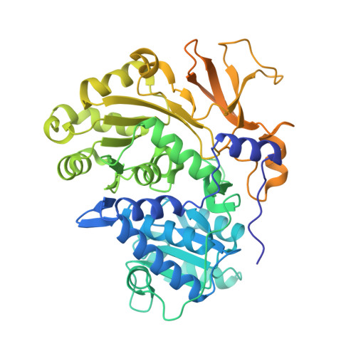
    Siderophores are small-molecule iron chelators produced by bacteria and other microorganisms for survival under iron limiting conditions such as found in a mammalian host. Siderophore biosynthesis is essential for the virulence of many important Gram-negative pathogens including Acinetobacter baumannii, Klebsiella pneumoniae, Pseudomonas aeruginosa, and Escherichia coli. We performed high-throughput screening against BasE, which is involved in siderophore biosynthesis in A. baumannii, and identified 6-phenyl-1-(pyridin-4-ylmethyl)-1H-pyrazolo[3,4-b]pyridine-4-carboxylic acid 15. Herein we report the synthesis, biochemical, and microbiological evaluation of a systematic series of analogues of the HTS hit 15. Analogue 67 is the most potent analogue with a KD of 2 nM against BasE. Structural characterization of the inhibitors with BasE reveals that they bind in a unique orientation in the active site, occupying all three substrate binding sites, and thus can be considered as multisubstrate inhibitors. These results provide a foundation for future studies aimed at increasing both enzyme potency and antibacterial activity.


  • Organizational Affiliation
    • Center for Drug Design, Academic Health Center, University of Minnesota , Minneapolis, Minnesota 55455, USA.

Macromolecule Content 

  • Total Structure Weight: 123.09 kDa 
  • Atom Count: 7,404 
  • Modeled Residue Count: 870 
  • Deposited Residue Count: 1,088 
  • Unique protein chains: 1

Macromolecules

Find similar proteins by:|  3D Structure
Entity ID: 1
MoleculeChains  Sequence LengthOrganismDetailsImage
Peptide arylation enzyme
A, B
544Acinetobacter baumannii ACICUMutation(s): 0 
Gene Names: ACICU_02578basE
EC: 6.2.1

Small Molecules

Ligands 4 Unique
IDChains Name / Formula / InChI Key2D Diagram3D Interactions
H89

Query on H89


Download:Ideal Coordinates CCD File
C [auth A],
I [auth B]
6-[4-(benzyloxy)phenyl]-1-(pyridin-4-ylmethyl)-1H-pyrazolo[3,4-b]pyridine-4-carboxylic acid
C26 H20 N4 O3
LEKPQTJLNDALBT-UHFFFAOYSA-N
MPD

Query on MPD


Download:Ideal Coordinates CCD File
H [auth A](4S)-2-METHYL-2,4-PENTANEDIOL
C6 H14 O2
SVTBMSDMJJWYQN-YFKPBYRVSA-N
MRD

Query on MRD


Download:Ideal Coordinates CCD File
L [auth B](4R)-2-METHYLPENTANE-2,4-DIOL
C6 H14 O2
SVTBMSDMJJWYQN-RXMQYKEDSA-N
CA

Query on CA


Download:Ideal Coordinates CCD File
D [auth A]
E [auth A]
F [auth A]
G [auth A]
J [auth B]
D [auth A],
E [auth A],
F [auth A],
G [auth A],
J [auth B],
K [auth B]
CALCIUM ION
Ca
BHPQYMZQTOCNFJ-UHFFFAOYSA-N

Experimental Data & Validation

Experimental Data

  • Method: X-RAY DIFFRACTION
  • Resolution: 2.10 Å
  • R-Value Free:  0.219 (Depositor), 0.220 (DCC) 
  • R-Value Work:  0.185 (Depositor) 
  • R-Value Observed: 0.187 (Depositor) 
Space Group: P 21 21 21
Unit Cell:
Length ( Å )Angle ( ˚ )
a = 66.093α = 90
b = 144.78β = 90
c = 148.675γ = 90
Software Package:
Software NamePurpose
PHENIXrefinement
REFMACrefinement
Blu-Icedata collection
HKL-2000data reduction
HKL-2000data scaling
REFMACphasing

Structure Validation

View Full Validation Report


Ligand Structure Quality Assessment 


Entry History 

Deposition Data

Revision History  (Full details and data files)

  • Version 1.0: 2012-10-03
    Type: Initial release
  • Version 1.1: 2019-07-17
    Changes: Data collection, Refinement description
  • Version 1.2: 2020-05-13
    Changes: Database references
  • Version 1.3: 2023-09-13
    Changes: Data collection, Database references, Derived calculations, Refinement description